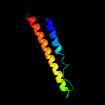
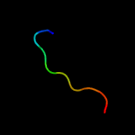
| 1 |
|
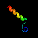
PDB 4xy3 chain A
Region: 49 - 115
Aligned: 67
Modelled: 67
Confidence: 31.4%
Identity: 18%
PDB header:protein transport
Chain: A: PDB Molecule:esx-1 secretion-associated protein espb;
PDBTitle: structure of esx-1 secreted protein espb
Phyre2
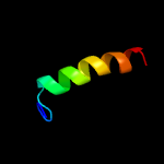
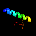
| 2 |
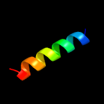
|
PDB 1wa8 chain A domain 1
Region: 40 - 63
Aligned: 24
Modelled: 24
Confidence: 31.0%
Identity: 29%
Fold: Ferritin-like
Superfamily: EsxAB dimer-like
Family: ESAT-6 like
Phyre2
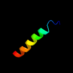
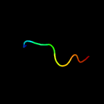
| 3 |
|
PDB 4iog chain D
Region: 40 - 63
Aligned: 24
Modelled: 24
Confidence: 28.6%
Identity: 33%
PDB header:unknown function
Chain: D: PDB Molecule:secreted protein esxb;
PDBTitle: the crystal structure of a secreted protein esxb (wild-type, in p212 space group) from bacillus anthracis str. sterne
Phyre2
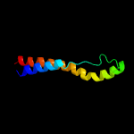
| 4 |
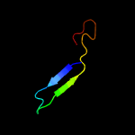
|
PDB 5xfs chain B
Region: 47 - 115
Aligned: 69
Modelled: 69
Confidence: 13.1%
Identity: 22%
PDB header:protein transport
Chain: B: PDB Molecule:ppe family protein ppe15;
PDBTitle: crystal structure of pe8-ppe15 in complex with espg5 from m.2 tuberculosis
Phyre2
| 5 |
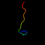
|
PDB 3gvm chain A
Region: 37 - 63
Aligned: 27
Modelled: 27
Confidence: 12.5%
Identity: 15%
PDB header:viral protein
Chain: A: PDB Molecule:putative uncharacterized protein sag1039;
PDBTitle: structure of the homodimeric wxg-100 family protein from streptococcus2 agalactiae
Phyre2
| 6 |
|
PDB 2vs0 chain B
Region: 43 - 63
Aligned: 21
Modelled: 19
Confidence: 12.1%
Identity: 5%
PDB header:cell invasion
Chain: B: PDB Molecule:virulence factor esxa;
PDBTitle: structural analysis of homodimeric staphylococcal aureus2 virulence factor esxa
Phyre2
| 7 |
|
PDB 3dzm chain B
Region: 49 - 76
Aligned: 28
Modelled: 28
Confidence: 11.6%
Identity: 21%
PDB header:unknown function
Chain: B: PDB Molecule:hypothetical conserved protein;
PDBTitle: crystal structure of a major outer membrane protein from thermus2 thermophilus hb27
Phyre2
| 8 |
|
PDB 1khb chain A domain 2
Region: 22 - 34
Aligned: 13
Modelled: 13
Confidence: 6.7%
Identity: 46%
Fold: PEP carboxykinase N-terminal domain
Superfamily: PEP carboxykinase N-terminal domain
Family: PEP carboxykinase N-terminal domain
Phyre2
| 9 |
|
PDB 1ic9 chain A
Region: 21 - 30
Aligned: 10
Modelled: 10
Confidence: 6.1%
Identity: 60%
PDB header:de novo protein
Chain: A: PDB Molecule:th10aox;
PDBTitle: nmr solution structure of the designed beta-sheet mini-2 protein th10aox
Phyre2
| 10 |
|
PDB 5cmo chain B
Region: 47 - 77
Aligned: 31
Modelled: 31
Confidence: 5.8%
Identity: 19%
PDB header:transferase
Chain: B: PDB Molecule:holo-[acyl-carrier-protein] synthase;
PDBTitle: crystal structure of holo-[acyl-carrier-protein] synthase (acps) from2 neisseria meningitidis
Phyre2
| 11 |
|
PDB 1icl chain A
Region: 21 - 30
Aligned: 10
Modelled: 10
Confidence: 5.6%
Identity: 60%
PDB header:de novo protein
Chain: A: PDB Molecule:th1ox;
PDBTitle: solution structure of designed beta-sheet mini-protein th1ox
Phyre2