Phyre Home Retrieve Phyre Job Id
Emailmdejesus@rockefeller.edu
DescriptionRVBD2601A_(-)_2930080_2930367
DateWed Aug 7 12:50:24 BST 2019
Unique Job ID11a84d385bea1788
SequenceMKTTLDLPDE ... Download FASTA
Job Typenormal
Job Expiry

Summary 

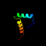
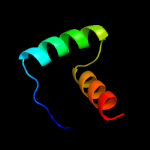
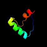
Top model
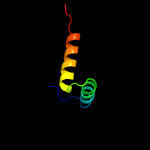
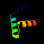
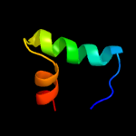
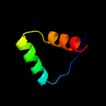
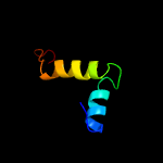
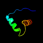
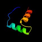
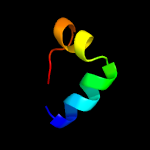
Image coloured by rainbow N → C terminus
Model dimensions (Å):X:39.727 Y:25.475 Z:26.196
Model (left) based on template d2bsqe1
Top template information
Fold:Ribbon-helix-helix
Superfamily:Ribbon-helix-helix
Family:Trafficking protein A-like
Confidence and coverage
Confidence: 91.9% Coverage: 45%
43 residues ( 45% of your sequence) have been modelled with 91.9% confidence by the single highest scoring template.
You may wish to submit your sequence to Phyrealarm. This will automatically scan your sequence every week for new potential templates as they appear in the Phyre2 library.
3D viewing
Interactive 3D view in JSmol
For other options to view your downloaded structure offline see the FAQ

Sequence analysis 

Download FASTA version

Secondary structure and disorder prediction [Show] 

Domain analysis [Show] 

Detailed template information [Hide] 


#
Template Alignment Coverage3D Model Confidence
% i.d. Template Information
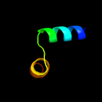
1d2bsqe1


91.9 21
Fold:Ribbon-helix-helix
Superfamily:Ribbon-helix-helix
Family:Trafficking protein A-like
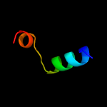
2c2h1oH_


90.9 21
PDB header:gene regulation/dna complex
Chain: H: PDB Molecule:trafficking protein a;
PDBTitle: structure of fitab bound to ir36 dna fragment
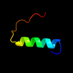
3c5x3tA_


88.5 41
PDB header:antitoxin/toxin
Chain: A: PDB Molecule:antitoxin vapb26;
PDBTitle: vapbc from mycobacterium tuberculosis
4c2an7A_


66.4 35
PDB header:dna binding protein
Chain: A: PDB Molecule:protein pard;
PDBTitle: solution structure of the bacterial antidote pard
5d1p94a_


58.5 24
Fold:Ribbon-helix-helix
Superfamily:Ribbon-helix-helix
Family:CopG-like
6c2k5jB_


57.4 21
PDB header:structural genomics, unknown function
Chain: B: PDB Molecule:uncharacterized protein yiif;
PDBTitle: solution structure of protein yiif from shigella flexneri2 serotype 5b (strain 8401) . northeast structural genomics3 consortium target sft1
7d1mnta_


50.1 23
Fold:Ribbon-helix-helix
Superfamily:Ribbon-helix-helix
Family:Arc/Mnt-like phage repressors
8c2ca9B_


45.0 18
PDB header:transcription
Chain: B: PDB Molecule:putative nickel-responsive regulator;
PDBTitle: apo-nikr from helicobacter pylori in closed trans-2 conformation
9d2hzab1


44.6 24
Fold:Ribbon-helix-helix
Superfamily:Ribbon-helix-helix
Family:CopG-like
10d2hzaa1


39.8 24
Fold:Ribbon-helix-helix
Superfamily:Ribbon-helix-helix
Family:CopG-like
11d2bj7a1


38.7 13
Fold:Ribbon-helix-helix
Superfamily:Ribbon-helix-helix
Family:CopG-like
12c2bj3D_


34.4 13
PDB header:transcription
Chain: D: PDB Molecule:nickel responsive regulator;
PDBTitle: nikr-apo
13c1q5vB_


30.6 24
PDB header:transcription
Chain: B: PDB Molecule:nickel responsive regulator;
PDBTitle: apo-nikr
14c2kj1A_


27.2 23
PDB header:transport protein
Chain: A: PDB Molecule:bm2 protein;
PDBTitle: cytoplasmic domain structure of bm2 proton channel from2 influenza b virus
15c3j9eD_


21.5 40
PDB header:viral protein
Chain: D: PDB Molecule:vp5;
PDBTitle: atomic structure of a non-enveloped virus reveals ph sensors for a2 coordinated process of cell entry
16c3qoqC_


20.8 23
PDB header:transcription/dna
Chain: C: PDB Molecule:alginate and motility regulator z;
PDBTitle: crystal structure of the transcription factor amrz in complex with the2 18 base pair amrz1 binding site
17c1rijA_


16.5 29
PDB header:de novo protein
Chain: A: PDB Molecule:e6apn1 peptide;
PDBTitle: e6-bind trp-cage (e6apn1)
18d1ujpa_


15.7 20
Fold:TIM beta/alpha-barrel
Superfamily:Ribulose-phoshate binding barrel
Family:Tryptophan biosynthesis enzymes
19c5tchG_


14.1 14
PDB header:lyase
Chain: G: PDB Molecule:tryptophan synthase alpha chain;
PDBTitle: crystal structure of tryptophan synthase from m. tuberculosis -2 ligand-free form, trpa-g66v mutant
20d1hw1a1


12.1 14
Fold:DNA/RNA-binding 3-helical bundle
Superfamily:"Winged helix" DNA-binding domain
Family:GntR-like transcriptional regulators
21c1u9pA_


not modelled 10.4 15
PDB header:unknown function
Chain: A: PDB Molecule:parc;
PDBTitle: permuted single-chain arc
22c5sxpG_


not modelled 9.1 70
PDB header:signaling protein/ligase
Chain: G: PDB Molecule:e3 ubiquitin-protein ligase itchy homolog;
PDBTitle: structural basis for the interaction between itch prr and beta-pix
23c5sxpF_


not modelled 9.0 70
PDB header:signaling protein/ligase
Chain: F: PDB Molecule:e3 ubiquitin-protein ligase itchy homolog;
PDBTitle: structural basis for the interaction between itch prr and beta-pix
24c5k9xA_


not modelled 9.0 20
PDB header:lyase
Chain: A: PDB Molecule:tryptophan synthase alpha chain;
PDBTitle: crystal structure of tryptophan synthase alpha chain from legionella2 pneumophila subsp. pneumophila
25c6g1nB_


not modelled 8.2 11
PDB header:antitoxin
Chain: B: PDB Molecule:antitoxin hicb;
PDBTitle: crystal structure of the burkholderia pseudomallei antitoxin hicb
26c6cmxA_


not modelled 8.1 13
PDB header:membrane protein
Chain: A: PDB Molecule:teneurin-2;
PDBTitle: human teneurin 2 extra-cellular region
27c2xa0A_


not modelled 7.6 29
PDB header:apoptosis
Chain: A: PDB Molecule:apoptosis regulator bcl-2;
PDBTitle: crystal structure of bcl-2 in complex with a bax bh32 peptide
28c6f1eA_


not modelled 6.8 7
PDB header:immune system
Chain: A: PDB Molecule:interferon gamma;
PDBTitle: crystal structure of olive flounder [paralichthys olivaceus]2 interferon gamma at 2.3 angstrom resolution
29c3fshC_


not modelled 6.3 33
PDB header:ligase
Chain: C: PDB Molecule:autocrine motility factor receptor, isoform 2;
PDBTitle: crystal structure of the ubiquitin conjugating enzyme2 ube2g2 bound to the g2br domain of ubiquitin ligase gp78
30c5j10A_


not modelled 5.7 46
PDB header:de novo protein
Chain: A: PDB Molecule:peptide design 2l4hc2_24;
PDBTitle: de novo design of protein homo-oligomers with modular hydrogen bond2 network-mediated specificity
31c4drvA_


not modelled 5.6 27
PDB header:viral protein
Chain: A: PDB Molecule:outer capsid protein vp4;
PDBTitle: cell attachment protein vp8* of a human rotavirus specifically2 interacts with a-type histo-blood group antigen
32d1j09a1


not modelled 5.4 24
Fold:An anticodon-binding domain of class I aminoacyl-tRNA synthetases
Superfamily:An anticodon-binding domain of class I aminoacyl-tRNA synthetases
Family:C-terminal domain of glutamyl-tRNA synthetase (GluRS)
33c5gj6B_


not modelled 5.4 36
PDB header:viral protein
Chain: B: PDB Molecule:outer capsid protein vp4;
PDBTitle: functional and structural characterization of p[19] rotavirus vp8*2 interaction with histo-blood group antigens
34c2lxpB_


not modelled 5.4 33
PDB header:ligase
Chain: B: PDB Molecule:e3 ubiquitin-protein ligase amfr;
PDBTitle: nmr structure of two domains in ubiquitin ligase gp78, ring and g2br,2 bound to its conjugating enzyme ube2g
35c6a7vU_


not modelled 5.3 28
PDB header:toxin/antitoxin
Chain: U: PDB Molecule:antitoxin vapb11;
PDBTitle: crystal structure of mycobacterium tuberculosis vapbc11 toxin-2 antitoxin complex
36c3h8kB_


not modelled 5.3 33
PDB header:ligase
Chain: B: PDB Molecule:autocrine motility factor receptor, isoform 2;
PDBTitle: crystal structure of ube2g2 complxed with the g2br domain of gp78 at2 1.8-a resolution
37c3pmdA_


not modelled 5.3 20
PDB header:lipid binding protein
Chain: A: PDB Molecule:conserved domain protein;
PDBTitle: crystal structure of the sporulation inhibitor pxo1-118 from bacillus2 anthracis
38c5ey5A_


not modelled 5.2 17
PDB header:lyase
Chain: A: PDB Molecule:lbcats-a;
PDBTitle: lbcats
39c3navB_


not modelled 5.1 9
PDB header:lyase
Chain: B: PDB Molecule:tryptophan synthase alpha chain;
PDBTitle: crystal structure of an alpha subunit of tryptophan synthase from2 vibrio cholerae o1 biovar el tor str. n16961

Binding site prediction 

Due to computational demand, binding site predictions are not run for batch jobs

If you want to predict binding sites, please manually submit your model of choice to 3DLigandSite