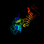
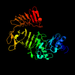
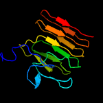
PDB header:protein transport Chain: A: PDB Molecule:pe family protein pe8; PDBTitle: crystal structure of pe8-ppe15 in complex with espg5 from m.2 tuberculosis
Confidence and coverage
Confidence:
99.8%
Coverage:
17%
77 residues ( 17% of your sequence) have been modelled with 99.8% confidence by the single highest scoring template.
Additional confident templates have been detected (see Domain analysis) which cover other regions of your sequence.
461 residues (100%) could be modelled at >90% confidence using multiple-templates.
You may wish to try resubmitting your sequence in "intensive" mode to model more of your sequence.
Warning: 82% of your sequence is predicted disordered. Disordered regions cannot be meaningfully predicted.
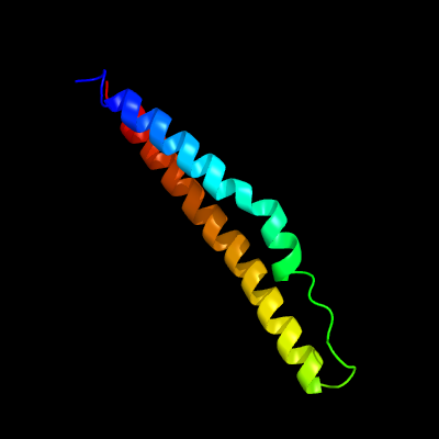
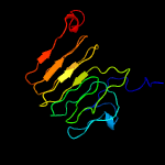
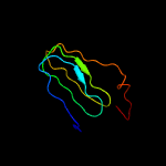
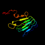
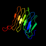
Region: 7 - 83 Aligned: 77 Modelled: 77 Confidence: 99.8% Identity: 42% PDB header:protein transport Chain: A: PDB Molecule:pe family protein pe8; PDBTitle: crystal structure of pe8-ppe15 in complex with espg5 from m.2 tuberculosis
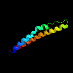
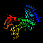
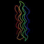
Region: 140 - 461 Aligned: 322 Modelled: 322 Confidence: 99.3% Identity: 29% PDB header:structural protein/contractile protein Chain: A: PDB Molecule:collagen i alpha 1; PDBTitle: the structure of collagen type i. single type i collagen2 molecule: rigid refinment
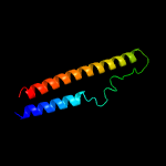
Region: 34 - 461 Aligned: 427 Modelled: 428 Confidence: 99.2% Identity: 23% PDB header:structural protein/contractile protein Chain: B: PDB Molecule:collagen i alpha 2; PDBTitle: the structure of collagen type i. single type i collagen2 molecule