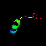
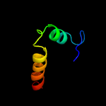
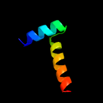
1 c4xgqD_
91.0
14
PDB header: toxin/antitoxinChain: D: PDB Molecule: antitoxin vapb30;PDBTitle: crystal structure of addiction module from mycobacterial species
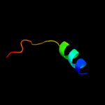
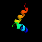
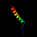
2 c4xgqB_
90.8
14
PDB header: toxin/antitoxinChain: B: PDB Molecule: antitoxin vapb30;PDBTitle: crystal structure of addiction module from mycobacterial species
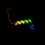
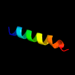
3 c4xgrH_
74.8
19
PDB header: toxin/antitoxinChain: H: PDB Molecule: antitoxin vapb30;PDBTitle: crystal structure of addiction module from mycobacterial species
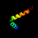
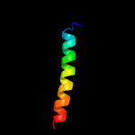
4 c4xgrF_
74.7
19
PDB header: toxin/antitoxinChain: F: PDB Molecule: antitoxin vapb30;PDBTitle: crystal structure of addiction module from mycobacterial species
5 c2k5jB_
65.3
11
PDB header: structural genomics, unknown functionChain: B: PDB Molecule: uncharacterized protein yiif;PDBTitle: solution structure of protein yiif from shigella flexneri2 serotype 5b (strain 8401) . northeast structural genomics3 consortium target sft1
6 c6gtsC_
57.2
15
PDB header: transcriptionChain: C: PDB Molecule: duf1778 domain-containing protein;PDBTitle: structure of the atat-atar complex bound dna
7 c6ajnF_
54.3
15
PDB header: toxinChain: F: PDB Molecule: duf1778 domain-containing protein;PDBTitle: crystal structure of atatr bound with accoa
8 d1y9ba1
42.9
18
Fold: Ribbon-helix-helixSuperfamily: Ribbon-helix-helixFamily: VCA0319-like
9 c2kelB_
37.6
33
PDB header: transcription repressorChain: B: PDB Molecule: uncharacterized protein 56b;PDBTitle: structure of the transcription regulator svtr from the2 hyperthermophilic archaeal virus sirv1
10 c2q2kA_
24.0
23
PDB header: dna binding protein/dnaChain: A: PDB Molecule: hypothetical protein;PDBTitle: structure of nucleic-acid binding protein
11 c2q2kB_
21.1
24
PDB header: dna binding protein/dnaChain: B: PDB Molecule: hypothetical protein;PDBTitle: structure of nucleic-acid binding protein
12 c6a7vU_
17.3
34
PDB header: toxin/antitoxinChain: U: PDB Molecule: antitoxin vapb11;PDBTitle: crystal structure of mycobacterium tuberculosis vapbc11 toxin-2 antitoxin complex
13 c3eabK_
16.2
35
PDB header: cell cycleChain: K: PDB Molecule: chmp1b;PDBTitle: crystal structure of spastin mit in complex with escrt iii
14 c3mx6A_
13.6
7
PDB header: hydrolaseChain: A: PDB Molecule: methionine aminopeptidase;PDBTitle: crystal structure of methionine aminopeptidase from rickettsia2 prowazekii bound to methionine
15 d2gg2a1
12.7
12
Fold: Creatinase/aminopeptidaseSuperfamily: Creatinase/aminopeptidaseFamily: Creatinase/aminopeptidase
16 d1u9pa1
12.2
21
Fold: Ribbon-helix-helixSuperfamily: Ribbon-helix-helixFamily: Arc/Mnt-like phage repressors
17 c4fo7B_
12.0
11
PDB header: hydrolaseChain: B: PDB Molecule: methionine aminopeptidase;PDBTitle: pseudomonas aeruginosa metap, in mn form
18 d1myka_
10.7
21
Fold: Ribbon-helix-helixSuperfamily: Ribbon-helix-helixFamily: Arc/Mnt-like phage repressors
19 d1bdta_
10.5
21
Fold: Ribbon-helix-helixSuperfamily: Ribbon-helix-helixFamily: Arc/Mnt-like phage repressors
20 c3tavA_
10.0
6
PDB header: hydrolaseChain: A: PDB Molecule: methionine aminopeptidase;PDBTitle: crystal structure of a methionine aminopeptidase from mycobacterium2 abscessus
21 d1baza_
not modelled
9.7
21
Fold: Ribbon-helix-helixSuperfamily: Ribbon-helix-helixFamily: Arc/Mnt-like phage repressors
22 c3f0nB_
not modelled
9.5
19
PDB header: lyaseChain: B: PDB Molecule: mevalonate pyrophosphate decarboxylase;PDBTitle: mus musculus mevalonate pyrophosphate decarboxylase
23 c5ca8A_
not modelled
9.4
38
PDB header: hydrolaseChain: A: PDB Molecule: protein sey1;PDBTitle: structures of the yeast dynamin-like gtpase sey1p in complex with gdp
24 d1myla_
not modelled
9.3
21
Fold: Ribbon-helix-helixSuperfamily: Ribbon-helix-helixFamily: Arc/Mnt-like phage repressors
25 c2k9iB_
not modelled
9.3
14
PDB header: dna binding proteinChain: B: PDB Molecule: uncharacterized protein orf56;PDBTitle: nmr structure of plasmid copy control protein orf56 from sulfolobus2 islandicus
26 d1p94a_
not modelled
9.1
27
Fold: Ribbon-helix-helixSuperfamily: Ribbon-helix-helixFamily: CopG-like
27 c3um2E_
not modelled
9.0
56
PDB header: membrane protein/transport proteinChain: E: PDB Molecule: charged multivesicular body protein 5;PDBTitle: crystal structure of the brox bro1 domain in complex with the c-2 terminal tail of chmp5
28 c3um1E_
not modelled
9.0
56
PDB header: membrane protein/transport proteinChain: E: PDB Molecule: charged multivesicular body protein 5;PDBTitle: crystal structure of the brox bro1 domain in complex with the c-2 terminal tail of chmp5
29 c3um2B_
not modelled
9.0
56
PDB header: membrane protein/transport proteinChain: B: PDB Molecule: charged multivesicular body protein 5;PDBTitle: crystal structure of the brox bro1 domain in complex with the c-2 terminal tail of chmp5
30 c3um0B_
not modelled
9.0
56
PDB header: membrane protein/transport proteinChain: B: PDB Molecule: charged multivesicular body protein 5;PDBTitle: crystal structure of the brox bro1 domain in complex with the c-2 terminal tail of chmp5
31 d1o0xa_
not modelled
8.9
8
Fold: Creatinase/aminopeptidaseSuperfamily: Creatinase/aminopeptidaseFamily: Creatinase/aminopeptidase
32 d1q9ja2
not modelled
8.2
20
Fold: CoA-dependent acyltransferasesSuperfamily: CoA-dependent acyltransferasesFamily: NRPS condensation domain (amide synthase)
33 d2hzaa1
not modelled
8.1
14
Fold: Ribbon-helix-helixSuperfamily: Ribbon-helix-helixFamily: CopG-like
34 d2a4da1
not modelled
7.9
28
Fold: UBC-likeSuperfamily: UBC-likeFamily: UBC-related
35 c3hxxA_
not modelled
7.8
18
PDB header: ligaseChain: A: PDB Molecule: alanyl-trna synthetase;PDBTitle: crystal structure of catalytic fragment of e. coli alars in complex2 with amppcp
36 d1cuka1
not modelled
7.7
21
Fold: RuvA C-terminal domain-likeSuperfamily: DNA helicase RuvA subunit, C-terminal domainFamily: DNA helicase RuvA subunit, C-terminal domain
37 c4egcA_
not modelled
7.6
50
PDB header: transcription/hydrolaseChain: A: PDB Molecule: maltose-binding periplasmic protein, homeobox protein six1PDBTitle: crystal structure of mbp-fused human six1 bound to human eya2 eya2 domain
38 c5h64b_
not modelled
7.3
16
PDB header: transferaseChain: B: PDB Molecule: regulatory-associated protein of mtor;PDBTitle: cryo-em structure of mtorc1
39 c2g6pA_
not modelled
7.2
11
PDB header: hydrolaseChain: A: PDB Molecule: methionine aminopeptidase 1;PDBTitle: crystal structure of truncated (delta 1-89) human methionine2 aminopeptidase type 1 in complex with pyridyl pyrimidine derivative
40 c1wkbA_
not modelled
6.9
25
PDB header: ligaseChain: A: PDB Molecule: leucyl-trna synthetase;PDBTitle: crystal structure of leucyl-trna synthetase from the2 archaeon pyrococcus horikoshii reveals a novel editing3 domain orientation
41 d1efva2
not modelled
6.9
28
Fold: DHS-like NAD/FAD-binding domainSuperfamily: DHS-like NAD/FAD-binding domainFamily: C-terminal domain of the electron transfer flavoprotein alpha subunit
42 c5vgtA_
not modelled
6.8
64
PDB header: viral proteinChain: A: PDB Molecule: gene 7 protein;PDBTitle: x-ray structure of bacteriophage sf6 tail adaptor protein gp7
43 c1zvvA_
not modelled
6.5
16
PDB header: transcription/dnaChain: A: PDB Molecule: glucose-resistance amylase regulator;PDBTitle: crystal structure of a ccpa-crh-dna complex
44 c3m92B_
not modelled
6.4
67
PDB header: structural genomics, unknown functionChain: B: PDB Molecule: protein ycin;PDBTitle: the structure of ycin, an unchracterized protein from shigella2 flexneri.
45 d1mylb_
not modelled
6.1
22
Fold: Ribbon-helix-helixSuperfamily: Ribbon-helix-helixFamily: Arc/Mnt-like phage repressors
46 c2gz5A_
not modelled
5.9
11
PDB header: hydrolaseChain: A: PDB Molecule: methionine aminopeptidase 1;PDBTitle: human type 1 methionine aminopeptidase in complex with ovalicin at 1.12 ang
47 c6n10A_
not modelled
5.9
19
PDB header: lyaseChain: A: PDB Molecule: diphosphomevalonate decarboxylase mvd1, peroxisomal;PDBTitle: crystal structure of arabidopsis thaliana mevalonate 5-diphosphate2 decarboxylase 1 complexed with (r)-mvapp
48 c4ifdK_
not modelled
5.7
37
PDB header: hydrolase/rnaChain: K: PDB Molecule: exosome complex exonuclease rrp6;PDBTitle: crystal structure of an 11-subunit eukaryotic exosome complex bound to2 rna
49 c4ariA_
not modelled
5.7
50
PDB header: ligase/rnaChain: A: PDB Molecule: leucine--trna ligase;PDBTitle: ternary complex of e. coli leucyl-trna synthetase, trna(leu) and the2 benzoxaborole an2679 in the editing conformation
50 d1bazb_
not modelled
5.7
22
Fold: Ribbon-helix-helixSuperfamily: Ribbon-helix-helixFamily: Arc/Mnt-like phage repressors
51 d1efpa2
not modelled
5.6
33
Fold: DHS-like NAD/FAD-binding domainSuperfamily: DHS-like NAD/FAD-binding domainFamily: C-terminal domain of the electron transfer flavoprotein alpha subunit
52 c3o0lB_
not modelled
5.3
43
PDB header: structural genomics, unknown functionChain: B: PDB Molecule: uncharacterized protein;PDBTitle: crystal structure of a pfam duf1425 family member (shew_1734) from2 shewanella sp. pv-4 at 1.81 a resolution