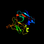
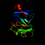
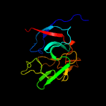
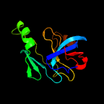
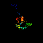
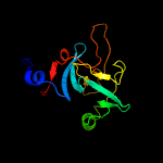
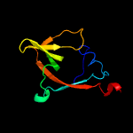
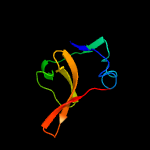
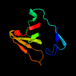
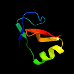
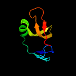
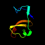
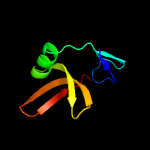
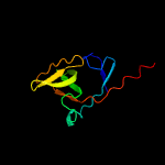
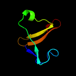
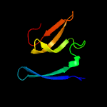
1 c4wviA_
100.0
37
PDB header: hydrolaseChain: A: PDB Molecule: maltose-binding periplasmic protein,signal peptidase ib;PDBTitle: crystal structure of the type-i signal peptidase from staphylococcus2 aureus (spsb) in complex with a substrate peptide (pep2).
2 d1b12a_
100.0
36
Fold: LexA/Signal peptidaseSuperfamily: LexA/Signal peptidaseFamily: Type 1 signal peptidase
3 c4n31A_
100.0
27
PDB header: cell adhesionChain: A: PDB Molecule: sipa;PDBTitle: structure and activity of streptococcus pyogenes sipa: a signal2 peptidase homologue essential for pilus polymerisation
4 c4me8A_
100.0
39
PDB header: hydrolaseChain: A: PDB Molecule: signal peptidase i;PDBTitle: crystal structure of a signal peptidase i (ef3073) from enterococcus2 faecalis v583 at 2.27 a resolution
5 c4nv4A_
100.0
37
PDB header: hydrolaseChain: A: PDB Molecule: signal peptidase i;PDBTitle: 1.8 angstrom crystal structure of signal peptidase i from bacillus2 anthracis.
6 c4k8wA_
100.0
28
PDB header: cell adhesionChain: A: PDB Molecule: lepa;PDBTitle: an arm-swapped dimer of the s. pyogenes pilin specific assembly factor2 sipa
7 d1f39a_
97.7
30
Fold: LexA/Signal peptidaseSuperfamily: LexA/Signal peptidaseFamily: LexA-related
8 c6a2rD_
97.4
20
PDB header: hydrolaseChain: D: PDB Molecule: lexa repressor;PDBTitle: mycobacterium tuberculosis lexa c-domain ii
9 c3k2zA_
97.4
28
PDB header: hydrolaseChain: A: PDB Molecule: lexa repressor;PDBTitle: crystal structure of a lexa protein from thermotoga maritima
10 d1umua_
96.9
28
Fold: LexA/Signal peptidaseSuperfamily: LexA/Signal peptidaseFamily: LexA-related
11 d1jhfa2
96.9
31
Fold: LexA/Signal peptidaseSuperfamily: LexA/Signal peptidaseFamily: LexA-related
12 c3jsoA_
96.8
30
PDB header: hydrolase/dnaChain: A: PDB Molecule: lexa repressor;PDBTitle: classic protein with a new twist: crystal structure of a lexa2 repressor dna complex
13 c1jhhB_
96.8
29
PDB header: hydrolaseChain: B: PDB Molecule: lexa repressor;PDBTitle: lexa s119a mutant
14 c3bdnB_
96.2
30
PDB header: transcription/dnaChain: B: PDB Molecule: lambda repressor;PDBTitle: crystal structure of the lambda repressor
15 c2hnfA_
96.0
25
PDB header: viral proteinChain: A: PDB Molecule: repressor protein ci101-229dm-k192a;PDBTitle: structure of a hyper-cleavable monomeric fragment of phage2 lambda repressor containing the cleavage site region
16 c2fjrB_
92.8
7
PDB header: transcription regulatorChain: B: PDB Molecule: repressor protein ci;PDBTitle: crystal structure of bacteriophage 186
17 d1hr0w_
60.5
32
Fold: OB-foldSuperfamily: Nucleic acid-binding proteinsFamily: Cold shock DNA-binding domain-like
18 c6c00A_
59.1
23
PDB header: translationChain: A: PDB Molecule: translation initiation factor if-1;PDBTitle: solution structure of translation initiation factor 1 from clostridium2 difficile
19 c2nchA_
58.7
29
PDB header: translationChain: A: PDB Molecule: translation initiation factor if-1;PDBTitle: solution structure of translation initiation factor if1 from wolbachia2 endosymbiont strain trs of brugia malayi
20 c4ql5A_
56.9
27
PDB header: translationChain: A: PDB Molecule: translation initiation factor if-1;PDBTitle: crystal structure of translation initiation factor if-1 from2 streptococcus pneumoniae tigr4
21 c3pbiA_
not modelled
49.8
12
PDB header: hydrolaseChain: A: PDB Molecule: invasion protein;PDBTitle: structure of the peptidoglycan hydrolase ripb (rv1478) from2 mycobacterium tuberculosis at 1.6 resolution
22 c3i4oA_
not modelled
49.7
23
PDB header: translationChain: A: PDB Molecule: translation initiation factor if-1;PDBTitle: crystal structure of translation initiation factor 1 from2 mycobacterium tuberculosis
23 d1ah9a_
not modelled
48.2
23
Fold: OB-foldSuperfamily: Nucleic acid-binding proteinsFamily: Cold shock DNA-binding domain-like
24 c4dqaA_
not modelled
45.7
17
PDB header: structural genomics, unknown functionChain: A: PDB Molecule: uncharacterized protein;PDBTitle: crystal structure of a putative carbohydrate binding protein2 (bacova_03559) from bacteroides ovatus atcc 8483 at 1.50 a resolution
25 c4bpp0_
not modelled
40.0
18
PDB header: ribosomeChain: 0: PDB Molecule: translation initiation factor eif-1a family protein;PDBTitle: the crystal structure of the eukaryotic 40s ribosomal subunit in2 complex with eif1 and eif1a - complex 4
26 c3j81i_
not modelled
39.6
32
PDB header: ribosomeChain: I: PDB Molecule: es8;PDBTitle: cryoem structure of a partial yeast 48s preinitiation complex
27 c2kogA_
not modelled
37.9
4
PDB header: membrane proteinChain: A: PDB Molecule: vesicle-associated membrane protein 2;PDBTitle: lipid-bound synaptobrevin solution nmr structure
28 d1v54d_
not modelled
37.4
10
Fold: Single transmembrane helixSuperfamily: Mitochondrial cytochrome c oxidase subunit IVFamily: Mitochondrial cytochrome c oxidase subunit IV
29 c2y69Q_
not modelled
36.9
10
PDB header: electron transportChain: Q: PDB Molecule: cytochrome c oxidase subunit 4 isoform 1;PDBTitle: bovine heart cytochrome c oxidase re-refined with molecular oxygen
30 c3hd7A_
not modelled
36.8
6
PDB header: exocytosisChain: A: PDB Molecule: vesicle-associated membrane protein 2;PDBTitle: helical extension of the neuronal snare complex into the membrane,2 spacegroup c 1 2 1
31 c2xivA_
not modelled
35.3
18
PDB header: structural proteinChain: A: PDB Molecule: hypothetical invasion protein;PDBTitle: structure of rv1477, hypothetical invasion protein of mycobacterium2 tuberculosis
32 d1jz8a5
not modelled
31.1
38
Fold: TIM beta/alpha-barrelSuperfamily: (Trans)glycosidasesFamily: beta-glycanases
33 d2ftxa1
not modelled
30.4
36
Fold: Kinetochore globular domain-likeSuperfamily: Kinetochore globular domainFamily: Spc25-like
34 c2oqkA_
not modelled
30.4
32
PDB header: translationChain: A: PDB Molecule: putative translation initiation factor eif-1a;PDBTitle: crystal structure of putative cryptosporidium parvum translation2 initiation factor eif-1a
35 d1bhga2
not modelled
29.4
24
Fold: Galactose-binding domain-likeSuperfamily: Galactose-binding domain-likeFamily: beta-Galactosidase/glucuronidase, N-terminal domain
36 c4hzbA_
not modelled
29.4
31
PDB header: hydrolaseChain: A: PDB Molecule: putative cytoplasmic protein;PDBTitle: crystal structure of the type vi semet effector-immunity complex tae3-2 tai3 from ralstonia pickettii
37 c2jv8A_
not modelled
26.6
38
PDB header: structural genomics, unknown functionChain: A: PDB Molecule: uncharacterized protein ne1242;PDBTitle: solution structure of protein ne1242 from nitrosomonas2 europaea. northeast structural genomics consortium target3 net4
38 d1jt8a_
not modelled
26.4
26
Fold: OB-foldSuperfamily: Nucleic acid-binding proteinsFamily: Cold shock DNA-binding domain-like
39 d1d7qa_
not modelled
25.6
32
Fold: OB-foldSuperfamily: Nucleic acid-binding proteinsFamily: Cold shock DNA-binding domain-like
40 c5td8D_
not modelled
23.8
36
PDB header: replicationChain: D: PDB Molecule: kinetochore protein spc25;PDBTitle: crystal structure of an extended dwarf ndc80 complex
41 d1knwa1
not modelled
23.5
14
Fold: Domain of alpha and beta subunits of F1 ATP synthase-likeSuperfamily: Alanine racemase C-terminal domain-likeFamily: Eukaryotic ODC-like
42 c3w5aC_
not modelled
23.3
13
PDB header: metal transport/membrane proteinChain: C: PDB Molecule: sarcolipin;PDBTitle: crystal structure of the calcium pump and sarcolipin from rabbit fast2 twitch skeletal muscle in the e1.mg2+ state
43 c4h1wB_
not modelled
23.3
13
PDB header: hydrolase/hydrolase regulatorChain: B: PDB Molecule: sarcolipin;PDBTitle: e1 structure of the (sr) ca2+-atpase in complex with sarcolipin
44 d1bhga3
not modelled
21.9
27
Fold: TIM beta/alpha-barrelSuperfamily: (Trans)glycosidasesFamily: beta-glycanases
45 d2ix0a2
not modelled
21.3
47
Fold: OB-foldSuperfamily: Nucleic acid-binding proteinsFamily: Cold shock DNA-binding domain-like
46 d1jnpa_
not modelled
20.2
18
Fold: Oncogene productsSuperfamily: Oncogene productsFamily: Oncogene products
47 d1zvpa1
not modelled
20.0
40
Fold: Ferredoxin-likeSuperfamily: ACT-likeFamily: VC0802-like
48 c4egxA_
not modelled
19.6
29
PDB header: transport proteinChain: A: PDB Molecule: kinesin-like protein kif1a;PDBTitle: crystal structure of kif1a cc1-fha tandem
49 c2vjjA_
not modelled
18.0
43
PDB header: viral proteinChain: A: PDB Molecule: tailspike protein;PDBTitle: tailspike protein of e.coli bacteriophage hk620 in complex with2 hexasaccharide
50 c4ejqB_
not modelled
17.7
29
PDB header: transport proteinChain: B: PDB Molecule: kinesin-like protein kif1a;PDBTitle: crystal structure of kif1a c-cc1-fha
51 d1a1xa_
not modelled
17.7
21
Fold: Oncogene productsSuperfamily: Oncogene productsFamily: Oncogene products
52 c2rtsA_
not modelled
16.8
32
PDB header: hydrolaseChain: A: PDB Molecule: chitinase;PDBTitle: chitin binding domain1
53 d1yq2a5
not modelled
16.8
33
Fold: TIM beta/alpha-barrelSuperfamily: (Trans)glycosidasesFamily: beta-glycanases
54 d1jsga_
not modelled
16.7
25
Fold: Oncogene productsSuperfamily: Oncogene productsFamily: Oncogene products
55 c1jlxB_
not modelled
16.6
32
PDB header: lectinChain: B: PDB Molecule: agglutinin;PDBTitle: agglutinin in complex with t-disaccharide
56 d2qmma1
not modelled
16.3
21
Fold: alpha/beta knotSuperfamily: alpha/beta knotFamily: AF1056-like
57 d2io8a2
not modelled
15.5
21
Fold: Cysteine proteinasesSuperfamily: Cysteine proteinasesFamily: CHAP domain
58 c3i86A_
not modelled
15.2
14
PDB header: hydrolaseChain: A: PDB Molecule: putative uncharacterized protein;PDBTitle: crystal structure of the p60 domain from m. avium subspecies2 paratuberculosis antigen map1204
59 c4mnoA_
not modelled
14.7
30
PDB header: translationChain: A: PDB Molecule: translation initiation factor 1a;PDBTitle: crystal structure of aif1a from pyrococcus abyssi
60 c2lzsE_
not modelled
14.4
33
PDB header: protein transportChain: E: PDB Molecule: sec-independent protein translocase protein tata;PDBTitle: tata oligomer
61 c3j1rQ_
not modelled
14.4
29
PDB header: cell adhesion, structural proteinChain: Q: PDB Molecule: archaeal adhesion filament core;PDBTitle: filaments from ignicoccus hospitalis show diversity of packing in2 proteins containing n-terminal type iv pilin helices
62 c3j1rR_
not modelled
14.4
29
PDB header: cell adhesion, structural proteinChain: R: PDB Molecule: archaeal adhesion filament core;PDBTitle: filaments from ignicoccus hospitalis show diversity of packing in2 proteins containing n-terminal type iv pilin helices
63 c3j1rH_
not modelled
14.4
29
PDB header: cell adhesion, structural proteinChain: H: PDB Molecule: archaeal adhesion filament core;PDBTitle: filaments from ignicoccus hospitalis show diversity of packing in2 proteins containing n-terminal type iv pilin helices
64 c3j1rD_
not modelled
14.4
29
PDB header: cell adhesion, structural proteinChain: D: PDB Molecule: archaeal adhesion filament core;PDBTitle: filaments from ignicoccus hospitalis show diversity of packing in2 proteins containing n-terminal type iv pilin helices
65 c3j1rM_
not modelled
14.4
29
PDB header: cell adhesion, structural proteinChain: M: PDB Molecule: archaeal adhesion filament core;PDBTitle: filaments from ignicoccus hospitalis show diversity of packing in2 proteins containing n-terminal type iv pilin helices
66 c3j1rB_
not modelled
14.4
29
PDB header: cell adhesion, structural proteinChain: B: PDB Molecule: archaeal adhesion filament core;PDBTitle: filaments from ignicoccus hospitalis show diversity of packing in2 proteins containing n-terminal type iv pilin helices
67 c3j1rK_
not modelled
14.4
29
PDB header: cell adhesion, structural proteinChain: K: PDB Molecule: archaeal adhesion filament core;PDBTitle: filaments from ignicoccus hospitalis show diversity of packing in2 proteins containing n-terminal type iv pilin helices
68 c3j1rG_
not modelled
14.4
29
PDB header: cell adhesion, structural proteinChain: G: PDB Molecule: archaeal adhesion filament core;PDBTitle: filaments from ignicoccus hospitalis show diversity of packing in2 proteins containing n-terminal type iv pilin helices
69 c3j1rN_
not modelled
14.4
29
PDB header: cell adhesion, structural proteinChain: N: PDB Molecule: archaeal adhesion filament core;PDBTitle: filaments from ignicoccus hospitalis show diversity of packing in2 proteins containing n-terminal type iv pilin helices
70 c3j1rE_
not modelled
14.4
29
PDB header: cell adhesion, structural proteinChain: E: PDB Molecule: archaeal adhesion filament core;PDBTitle: filaments from ignicoccus hospitalis show diversity of packing in2 proteins containing n-terminal type iv pilin helices
71 c3j1rO_
not modelled
14.4
29
PDB header: cell adhesion, structural proteinChain: O: PDB Molecule: archaeal adhesion filament core;PDBTitle: filaments from ignicoccus hospitalis show diversity of packing in2 proteins containing n-terminal type iv pilin helices
72 c3j1rI_
not modelled
14.4
29
PDB header: cell adhesion, structural proteinChain: I: PDB Molecule: archaeal adhesion filament core;PDBTitle: filaments from ignicoccus hospitalis show diversity of packing in2 proteins containing n-terminal type iv pilin helices
73 c3j1rF_
not modelled
14.4
29
PDB header: cell adhesion, structural proteinChain: F: PDB Molecule: archaeal adhesion filament core;PDBTitle: filaments from ignicoccus hospitalis show diversity of packing in2 proteins containing n-terminal type iv pilin helices
74 c3j1rT_
not modelled
14.4
29
PDB header: cell adhesion, structural proteinChain: T: PDB Molecule: archaeal adhesion filament core;PDBTitle: filaments from ignicoccus hospitalis show diversity of packing in2 proteins containing n-terminal type iv pilin helices
75 c3j1rL_
not modelled
14.4
29
PDB header: cell adhesion, structural proteinChain: L: PDB Molecule: archaeal adhesion filament core;PDBTitle: filaments from ignicoccus hospitalis show diversity of packing in2 proteins containing n-terminal type iv pilin helices
76 c3j1rA_
not modelled
14.4
29
PDB header: cell adhesion, structural proteinChain: A: PDB Molecule: archaeal adhesion filament core;PDBTitle: filaments from ignicoccus hospitalis show diversity of packing in2 proteins containing n-terminal type iv pilin helices
77 c3j1rP_
not modelled
14.4
29
PDB header: cell adhesion, structural proteinChain: P: PDB Molecule: archaeal adhesion filament core;PDBTitle: filaments from ignicoccus hospitalis show diversity of packing in2 proteins containing n-terminal type iv pilin helices
78 c3j1rC_
not modelled
14.4
29
PDB header: cell adhesion, structural proteinChain: C: PDB Molecule: archaeal adhesion filament core;PDBTitle: filaments from ignicoccus hospitalis show diversity of packing in2 proteins containing n-terminal type iv pilin helices
79 c3j1rU_
not modelled
14.4
29
PDB header: cell adhesion, structural proteinChain: U: PDB Molecule: archaeal adhesion filament core;PDBTitle: filaments from ignicoccus hospitalis show diversity of packing in2 proteins containing n-terminal type iv pilin helices
80 c3j1rJ_
not modelled
14.4
29
PDB header: cell adhesion, structural proteinChain: J: PDB Molecule: archaeal adhesion filament core;PDBTitle: filaments from ignicoccus hospitalis show diversity of packing in2 proteins containing n-terminal type iv pilin helices
81 c3j1rS_
not modelled
14.4
29
PDB header: cell adhesion, structural proteinChain: S: PDB Molecule: archaeal adhesion filament core;PDBTitle: filaments from ignicoccus hospitalis show diversity of packing in2 proteins containing n-terminal type iv pilin helices
82 c1jdmA_
not modelled
14.1
8
PDB header: membrane proteinChain: A: PDB Molecule: sarcolipin;PDBTitle: nmr structure of sarcolipin
83 c2pkpA_
not modelled
13.7
40
PDB header: lyaseChain: A: PDB Molecule: homoaconitase small subunit;PDBTitle: crystal structure of 3-isopropylmalate dehydratase (leud)2 from methhanocaldococcus jannaschii dsm2661 (mj1271)
84 c2na9A_
not modelled
13.5
22
PDB header: signaling proteinChain: A: PDB Molecule: cytokine receptor common subunit beta;PDBTitle: transmembrane structure of the p441a mutant of the cytokine receptor2 common subunit beta
85 c4wolA_
not modelled
13.4
16
PDB header: signaling proteinChain: A: PDB Molecule: tyro protein tyrosine kinase-binding protein;PDBTitle: crystal structure of the dap12 transmembrane domain in lipidic cubic2 phase
86 c2l34B_
not modelled
13.4
16
PDB header: protein bindingChain: B: PDB Molecule: tyro protein tyrosine kinase-binding protein;PDBTitle: structure of the dap12 transmembrane homodimer
87 c2pwyB_
not modelled
13.2
19
PDB header: transferaseChain: B: PDB Molecule: trna (adenine-n(1)-)-methyltransferase;PDBTitle: crystal structure of a m1a58 trna methyltransferase
88 c3mt1B_
not modelled
13.1
17
PDB header: lyaseChain: B: PDB Molecule: putative carboxynorspermidine decarboxylase protein;PDBTitle: crystal structure of putative carboxynorspermidine decarboxylase2 protein from sinorhizobium meliloti
89 c3nx6A_
not modelled
13.1
23
PDB header: chaperoneChain: A: PDB Molecule: 10kda chaperonin;PDBTitle: crystal structure of co-chaperonin, groes (xoo4289) from xanthomonas2 oryzae pv. oryzae kacc10331
90 c4d7zA_
not modelled
13.0
38
PDB header: lyaseChain: A: PDB Molecule: aspartate 1-decarboxylase beta chain;PDBTitle: e. coli l-aspartate-alpha-decarboxylase mutant n72q to a resolution of2 1.9 angstroms
91 c4wo1C_
not modelled
12.9
16
PDB header: signaling proteinChain: C: PDB Molecule: tyro protein tyrosine kinase-binding protein;PDBTitle: crystal structure of the dap12 transmembrane domain in lipid cubic2 phase
92 c4wo1A_
not modelled
12.9
16
PDB header: signaling proteinChain: A: PDB Molecule: tyro protein tyrosine kinase-binding protein;PDBTitle: crystal structure of the dap12 transmembrane domain in lipid cubic2 phase
93 c4wo1B_
not modelled
12.9
16
PDB header: signaling proteinChain: B: PDB Molecule: tyro protein tyrosine kinase-binding protein;PDBTitle: crystal structure of the dap12 transmembrane domain in lipid cubic2 phase
94 c4wo1D_
not modelled
12.9
16
PDB header: signaling proteinChain: D: PDB Molecule: tyro protein tyrosine kinase-binding protein;PDBTitle: crystal structure of the dap12 transmembrane domain in lipid cubic2 phase
95 c5djoB_
not modelled
12.9
24
PDB header: transport proteinChain: B: PDB Molecule: kinesin-like protein;PDBTitle: crystal structure of the cc1-fha tandem of kinesin-3 kif13a
96 c3n29A_
not modelled
12.8
15
PDB header: lyaseChain: A: PDB Molecule: carboxynorspermidine decarboxylase;PDBTitle: crystal structure of carboxynorspermidine decarboxylase complexed with2 norspermidine from campylobacter jejuni
97 c2mi2A_
not modelled
12.8
20
PDB header: transport proteinChain: A: PDB Molecule: sec-independent protein translocase protein tatb;PDBTitle: solution structure of the e. coli tatb protein in dpc micelles
98 c2pfuA_
not modelled
12.7
11
PDB header: transport proteinChain: A: PDB Molecule: biopolymer transport exbd protein;PDBTitle: nmr strcuture determination of the periplasmic domain of exbd from2 e.coli
99 c4wolB_
not modelled
12.4
16
PDB header: signaling proteinChain: B: PDB Molecule: tyro protein tyrosine kinase-binding protein;PDBTitle: crystal structure of the dap12 transmembrane domain in lipidic cubic2 phase


