Phyre Home Retrieve Phyre Job Id
Emailmdejesus@rockefeller.edu
DescriptionRVBD2964B_(RVBD2964B)_3318111_3318296
DateThu Aug 8 16:20:12 BST 2019
Unique Job ID5766ff8ba7ce43a8
SequenceMFTARIRALA ... Download FASTA
Job Typenormal
Job Expiry

Summary 

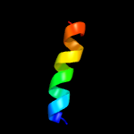
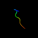
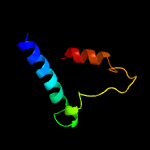
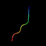
Top model
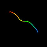
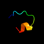
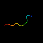
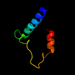
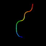
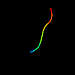
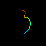
Image coloured by rainbow N → C terminus
Model dimensions (Å):X:12.628 Y:17.886 Z:24.901
Model (left) based on template c1hgvA_
Top template information
PDB header:virus
Chain: A: PDB Molecule:ph75 inovirus major coat protein;
PDBTitle: filamentous bacteriophage ph75
Confidence and coverage
Confidence: 21.3% Coverage: 28%
17 residues ( 28% of your sequence) have been modelled with 21.3% confidence by the single highest scoring template.
You may wish to submit your sequence to Phyrealarm. This will automatically scan your sequence every week for new potential templates as they appear in the Phyre2 library.
Please note: You must be registered and logged in to use Phyrealarm.
3D viewing
Interactive 3D view in JSmol
For other options to view your downloaded structure offline see the FAQ

Sequence analysis 

Download FASTA version

Secondary structure and disorder prediction [Show] 

Domain analysis [Show] 

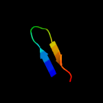
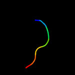
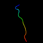
Detailed template information [Hide] 


#
Template Alignment Coverage3D Model Confidence
% i.d. Template Information
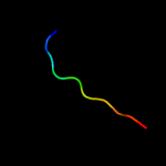
1c1hgvA_


21.3 59
PDB header:virus
Chain: A: PDB Molecule:ph75 inovirus major coat protein;
PDBTitle: filamentous bacteriophage ph75
2c5jyxB_


19.8 0
PDB header:transferase
Chain: B: PDB Molecule:archeaosine synthase quef-like;
PDBTitle: crystal structure of the covalent thioimide intermediate of the2 archaeosine synthase quef-like
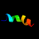
3c1hgzA_


15.8 59
PDB header:virus
Chain: A: PDB Molecule:ph75 inovirus major coat protein;
PDBTitle: filamentous bacteriophage ph75
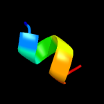
4c3zheC_


13.4 75
PDB header:mrna-binding protein
Chain: C: PDB Molecule:nonsense-mediated mrna decay protein;
PDBTitle: structure of the c. elegans smg5-smg7 complex
5d1xmwa1


11.6 50
Fold:Immunoglobulin-like beta-sandwich
Superfamily:Immunoglobulin
Family:I set domains
6d1c55a_


9.7 50
Fold:Knottins (small inhibitors, toxins, lectins)
Superfamily:Scorpion toxin-like
Family:Short-chain scorpion toxins
7c3oaiB_


8.8 40
PDB header:membrane protein, cell adhesion
Chain: B: PDB Molecule:maltose-binding periplasmic protein, myelin protein p0;
PDBTitle: crystal structure of the extra-cellular domain of human myelin protein2 zero
8c6idvB_


8.2 15
PDB header:ligase
Chain: B: PDB Molecule:peptide asparaginyl ligases;
PDBTitle: peptide asparaginyl ligases from viola yedoensis
9c3bp1A_


7.6 38
PDB header:oxidoreductase
Chain: A: PDB Molecule:nadph-dependent 7-cyano-7-deazaguanine reductase;
PDBTitle: crystal structure of putative 7-cyano-7-deazaguanine reductase quef2 from vibrio cholerae o1 biovar eltor
10d1wwbx_


7.6 43
Fold:Immunoglobulin-like beta-sandwich
Superfamily:Immunoglobulin
Family:I set domains
11c1kl8B_


7.5 71
PDB header:toxin
Chain: B: PDB Molecule:neuronal acetylcholine receptor protein, alpha-7
PDBTitle: nmr structural analysis of the complex formed between alpha-2 bungarotoxin and the principal alpha-neurotoxin binding3 sequence on the alpha7 subunit of a neuronal nicotinic4 acetylcholine receptor
12c1kc4B_


7.5 71
PDB header:toxin
Chain: B: PDB Molecule:neuronal acetylcholine receptor protein, alpha-7
PDBTitle: nmr structural analysis of the complex formed between alpha-2 bungarotoxin and the principal alpha-neurotoxin binding3 sequence on the alpha7 subunit of a neuronal nicotinic4 acetylcholine receptor
13c5h0iB_


7.3 15
PDB header:hydrolase
Chain: B: PDB Molecule:asparaginyl endopeptidase;
PDBTitle: name to be released when published
14d1xiwb_


6.8 36
Fold:Immunoglobulin-like beta-sandwich
Superfamily:Immunoglobulin
Family:I set domains
15c2vraD_


6.0 43
PDB header:cell adhesion
Chain: D: PDB Molecule:roundabout 1;
PDBTitle: drosophila robo ig1-2 (monoclinic form)
16c4f8bE_


6.0 40
PDB header:oxidoreductase
Chain: E: PDB Molecule:nadph-dependent 7-cyano-7-deazaguanine reductase;
PDBTitle: crystal structure of the covalent thioimide intermediate of unimodular2 nitrile reductase quef
17d1m4ka2


5.9 50
Fold:Immunoglobulin-like beta-sandwich
Superfamily:Immunoglobulin
Family:I set domains
18d1olla2


5.6 50
Fold:Immunoglobulin-like beta-sandwich
Superfamily:Immunoglobulin
Family:I set domains
19d1f97a1


5.5 57
Fold:Immunoglobulin-like beta-sandwich
Superfamily:Immunoglobulin
Family:V set domains (antibody variable domain-like)
20c1cdhA_


5.3 57
PDB header:t-cell surface glycoprotein
Chain: A: PDB Molecule:t cell surface glycoprotein cd4;
PDBTitle: structures of an hiv and mhc binding fragment from human cd4 as2 refined in two crystal lattices
21c3bp6B_


not modelled 5.3 14
PDB header:signaling protein
Chain: B: PDB Molecule:programmed cell death 1 ligand 2;
PDBTitle: crystal structure of the mouse pd-1 mutant and pd-l2 complex
22d1tnna_


not modelled 5.2 17
Fold:Immunoglobulin-like beta-sandwich
Superfamily:Immunoglobulin
Family:I set domains
23c3vh8H_


not modelled 5.2 36
PDB header:immune system
Chain: H: PDB Molecule:killer cell immunoglobulin-like receptor 3dl1;
PDBTitle: kir3dl1 in complex with hla-b*5701
24d1mvka_


not modelled 5.2 75
Fold:beta-Grasp (ubiquitin-like)
Superfamily:Immunoglobulin-binding domains
Family:Immunoglobulin-binding domains
25c3rbgB_


not modelled 5.2 43
PDB header:immune system
Chain: B: PDB Molecule:cytotoxic and regulatory t-cell molecule;
PDBTitle: crystal structure analysis of class-i mhc restricted t-cell associated2 molecule
26c4nikB_


not modelled 5.2 29
PDB header:oncoprotein/immune system
Chain: B: PDB Molecule:single-chain fv fragment antibody;
PDBTitle: structure of human gankyrin in complex to the single chain antibody f5
27d1nkra2


not modelled 5.1 50
Fold:Immunoglobulin-like beta-sandwich
Superfamily:Immunoglobulin
Family:I set domains
28d1ry7b1


not modelled 5.1 57
Fold:Immunoglobulin-like beta-sandwich
Superfamily:Immunoglobulin
Family:I set domains
29c2a38A_


not modelled 5.1 43
PDB header:structural protein
Chain: A: PDB Molecule:titin;
PDBTitle: crystal structure of the n-terminus of titin
30c6onbB_


not modelled 5.0 38
PDB header:cell adhesion
Chain: B: PDB Molecule:neuronal igcam-5;
PDBTitle: crystal structure of the zig-8-rig-5 ig1-ig1 heterodimer, monoclinic2 form

Binding site prediction 

Due to computational demand, binding site predictions are not run for batch jobs

If you want to predict binding sites, please manually submit your model of choice to 3DLigandSite