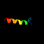
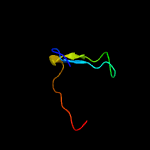
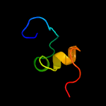
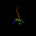
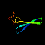
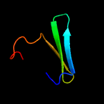
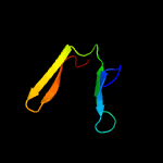
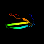
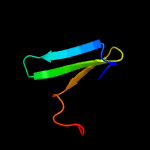
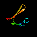
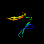
1 c2na7A_
60.8
37
PDB header: apoptosisChain: A: PDB Molecule: tumor necrosis factor receptor superfamily member 6;PDBTitle: transmembrane domain of human fas/cd95 death receptor
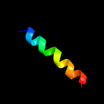
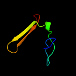
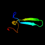
2 c2na7B_
57.1
37
PDB header: apoptosisChain: B: PDB Molecule: tumor necrosis factor receptor superfamily member 6;PDBTitle: transmembrane domain of human fas/cd95 death receptor
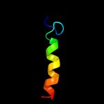
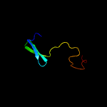
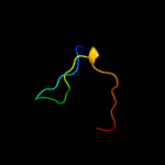
3 c2na7C_
57.1
37
PDB header: apoptosisChain: C: PDB Molecule: tumor necrosis factor receptor superfamily member 6;PDBTitle: transmembrane domain of human fas/cd95 death receptor
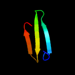
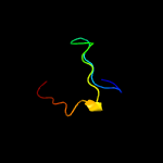
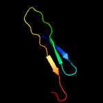
4 c3kifD_
53.6
25
PDB header: sugar binding proteinChain: D: PDB Molecule: 5-bladed beta-propeller lectin;PDBTitle: the crystal structures of two fragments truncated from 5-bladed beta-2 propeller lectin, tachylectin-2 (lib1-b7-18 and lib2-d2-15)
5 d1e5wa2
31.5
17
Fold: PH domain-like barrelSuperfamily: PH domain-likeFamily: Third domain of FERM
6 c4cvuA_
29.6
18
PDB header: hydrolaseChain: A: PDB Molecule: beta-mannosidase;PDBTitle: structure of fungal beta-mannosidase from glycoside hydrolase family 22 of trichoderma harzianum
7 d1j19a2
25.7
17
Fold: PH domain-like barrelSuperfamily: PH domain-likeFamily: Third domain of FERM
8 c3k8rA_
25.6
26
PDB header: structural genomics, unknown functionChain: A: PDB Molecule: uncharacterized protein;PDBTitle: crystal structure of protein of unknown function (yp_427503.1) from2 rhodospirillum rubrum atcc 11170 at 2.75 a resolution
9 d1tl2a_
23.1
17
Fold: 5-bladed beta-propellerSuperfamily: Tachylectin-2Family: Tachylectin-2
10 d1ef1a2
22.9
15
Fold: PH domain-like barrelSuperfamily: PH domain-likeFamily: Third domain of FERM
11 c3kihC_
22.7
14
PDB header: sugar binding proteinChain: C: PDB Molecule: 5-bladed beta-propeller lectin;PDBTitle: the crystal structures of two fragments truncated from 5-bladed beta-2 propeller lectin, tachylectin-2 (lib2-d2-15)
12 d1isna2
21.7
13
Fold: PH domain-like barrelSuperfamily: PH domain-likeFamily: Third domain of FERM
13 d1gg3a2
19.6
13
Fold: PH domain-like barrelSuperfamily: PH domain-likeFamily: Third domain of FERM
14 d2cx1a2
18.6
23
Fold: Cystatin-likeSuperfamily: Pre-PUA domainFamily: Hypothetical protein APE0525, N-terminal domain
15 c5c2mA_
18.3
15
PDB header: structural proteinChain: A: PDB Molecule: predicted protein;PDBTitle: the de novo evolutionary emergence of a symmetrical protein is shaped2 by folding constraints
16 d2zpya2
18.1
15
Fold: PH domain-like barrelSuperfamily: PH domain-likeFamily: Third domain of FERM
17 d1h4ra2
16.4
11
Fold: PH domain-like barrelSuperfamily: PH domain-likeFamily: Third domain of FERM
18 c2x8nA_
9.1
17
PDB header: structural genomicsChain: A: PDB Molecule: cv0863;PDBTitle: solution nmr structure of uncharacterized protein cv0863 from2 chromobacterium violaceum. northeast structural genomics target3 (nesg) target cvt3. ocsp target cv0863.
19 c2i1jA_
9.0
17
PDB header: cell adhesion, membrane proteinChain: A: PDB Molecule: moesin;PDBTitle: moesin from spodoptera frugiperda at 2.1 angstroms resolution
20 c3nqnB_
8.6
17
PDB header: structural genomics, unknown functionChain: B: PDB Molecule: uncharacterized protein;PDBTitle: crystal structure of a protein with unknown function. (dr_2006) from2 deinococcus radiodurans at 1.88 a resolution
21 c3osvC_
not modelled
8.4
28
PDB header: structural proteinChain: C: PDB Molecule: flagellar basal-body rod modification protein flgd;PDBTitle: the crytsal structure of flgd from p. aeruginosa
22 c2i1kA_
not modelled
7.6
17
PDB header: cell adhesion, membrane proteinChain: A: PDB Molecule: moesin;PDBTitle: moesin from spodoptera frugiperda reveals the coiled-coil domain at2 3.0 angstrom resolution
23 c4h7lB_
not modelled
7.4
16
PDB header: structural genomics, unknown functionChain: B: PDB Molecule: uncharacterized protein;PDBTitle: crystal structure of plim_4148 protein from planctomyces limnophilus
24 c6d2qA_
not modelled
7.2
21
PDB header: signaling proteinChain: A: PDB Molecule: ferm, rhogef (arhgef) and pleckstrin domain protein 1PDBTitle: crystal structure of the ferm domain of zebrafish farp1
25 c2lnzA_
not modelled
7.1
60
PDB header: protein bindingChain: A: PDB Molecule: ubiquitin-like protein mdy2;PDBTitle: solution structure of the get5 carboxyl domain from s. cerevisiae
26 c3vejB_
not modelled
7.0
60
PDB header: protein bindingChain: B: PDB Molecule: ubiquitin-like protein mdy2;PDBTitle: crystal structure of the get5 carboxyl domain from s. cerevisiae
27 c1e5wA_
not modelled
6.8
15
PDB header: membrane proteinChain: A: PDB Molecule: moesin;PDBTitle: structure of isolated ferm domain and first long helix of moesin
28 d1hnga1
not modelled
6.6
37
Fold: Immunoglobulin-like beta-sandwichSuperfamily: ImmunoglobulinFamily: V set domains (antibody variable domain-like)
29 d1rl6a2
not modelled
6.1
57
Fold: Ribosomal protein L6Superfamily: Ribosomal protein L6Family: Ribosomal protein L6
30 d2zjre1
not modelled
6.0
43
Fold: Ribosomal protein L6Superfamily: Ribosomal protein L6Family: Ribosomal protein L6
31 d2diga1
not modelled
5.7
32
Fold: SH3-like barrelSuperfamily: Tudor/PWWP/MBTFamily: Tudor domain
32 d2j01h2
not modelled
5.6
43
Fold: Ribosomal protein L6Superfamily: Ribosomal protein L6Family: Ribosomal protein L6
33 c2yowB_
not modelled
5.6
9
PDB header: oxidoreductaseChain: B: PDB Molecule: rbam17540;PDBTitle: bacillus amyloliquefaciens cbm33
34 c2e6xD_
not modelled
5.5
50
PDB header: structural genomics, unknown functionChain: D: PDB Molecule: hypothetical protein ttha1281;PDBTitle: x-ray structure of tt1592 from thermus thermophilus hb8
35 c2digA_
not modelled
5.5
32
PDB header: dna binding proteinChain: A: PDB Molecule: lamin-b receptor;PDBTitle: solusion structure of the todor domain of human lamin-b2 receptor
36 c3c8lB_
not modelled
5.4
25
PDB header: unknown functionChain: B: PDB Molecule: ftsz-like protein of unknown function;PDBTitle: crystal structure of a ftsz-like protein of unknown function2 (npun_r1471) from nostoc punctiforme pcc 73102 at 1.22 a resolution
37 c1u2pA_
not modelled
5.4
50
PDB header: hydrolaseChain: A: PDB Molecule: low molecular weight protein-tyrosine-PDBTitle: crystal structure of mycobacterium tuberculosis low2 molecular protein tyrosine phosphatase (mptpa) at 1.9a3 resolution
38 d1dg9a_
not modelled
5.4
18
Fold: Phosphotyrosine protein phosphatases I-likeSuperfamily: Phosphotyrosine protein phosphatases IFamily: Low-molecular-weight phosphotyrosine protein phosphatases
39 d2qamg2
not modelled
5.2
57
Fold: Ribosomal protein L6Superfamily: Ribosomal protein L6Family: Ribosomal protein L6
40 c4a02A_
not modelled
5.2
9
PDB header: chitin binding proteinChain: A: PDB Molecule: chitin binding protein;PDBTitle: x-ray crystallographic structure of efcbm33a


