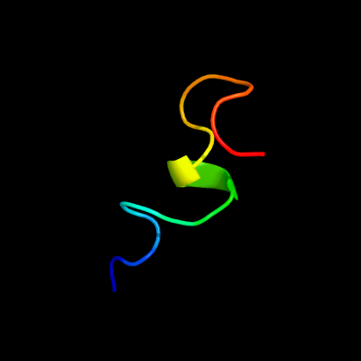
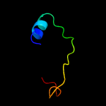
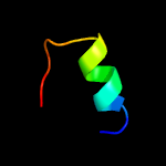
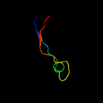
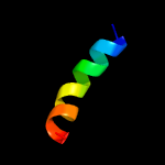
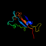
PDB header:viral protein Chain: A: PDB Molecule:envelope glycoprotein; PDBTitle: structure of the ebola virus envelope protein mper/tm domain and its2 interaction with the fusion loop explains their fusion activity
Confidence and coverage
Confidence:
41.6%
Coverage:
9%
19 residues ( 9% of your sequence) have been modelled with 41.6% confidence by the single highest scoring template.
You may wish to submit your sequence to Phyrealarm. This will automatically scan your sequence every week for new potential templates as they appear in the Phyre2 library.
Please note: You must be registered and logged in to use Phyrealarm.
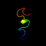
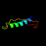
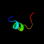
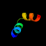
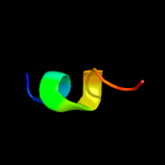
Region: 144 - 175 Aligned: 32 Modelled: 32 Confidence: 34.3% Identity: 22% PDB header:hydrolase Chain: A: PDB Molecule:chromodomain-helicase-dna-binding protein 7; PDBTitle: solution structures of the brk domains of the human chromo2 helicase domain 7 and 8, reveals structural similarity3 with gyf domain suggesting a role in protein interaction
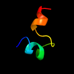
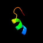
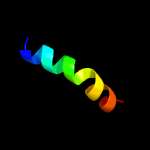
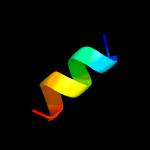
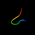
Region: 80 - 125 Aligned: 43 Modelled: 46 Confidence: 30.2% Identity: 23% PDB header:unknown function Chain: B: PDB Molecule:duf269-containing protein; PDBTitle: crystal structure of cce_0566 from the cyanobacterium cyanothece2 51142, a protein associated with nitrogen fixation from the duf2693 family
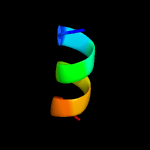
Region: 144 - 159 Aligned: 16 Modelled: 16 Confidence: 23.2% Identity: 38% PDB header:hydrolase Chain: A: PDB Molecule:chromodomain-helicase-dna-binding protein 8; PDBTitle: solution structures of the brk domains of the human chromo2 helicase domain 7 and 8, reveals structural similarity3 with gyf domain suggesting a role in protein interaction