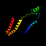
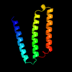
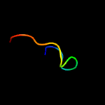
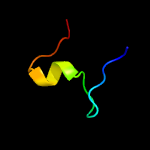
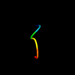
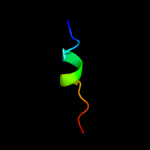
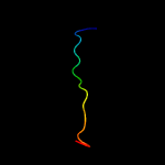
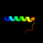
1 c6humC_
100.0
37
PDB header: proton transportChain: C: PDB Molecule: nad(p)h-quinone oxidoreductase subunit 3;PDBTitle: structure of the photosynthetic complex i from thermosynechococcus2 elongatus
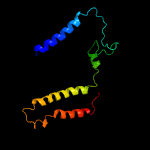
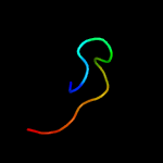
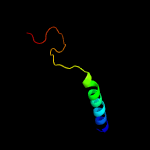
2 c5lc5A_
100.0
28
PDB header: oxidoreductaseChain: A: PDB Molecule: nadh-ubiquinone oxidoreductase chain 3;PDBTitle: structure of mammalian respiratory complex i, class2
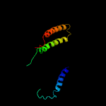
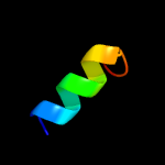
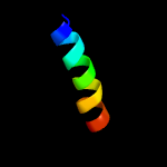
3 c6gcs3_
100.0
20
PDB header: oxidoreductaseChain: 3: PDB Molecule: nd3 subunit (nu3m);PDBTitle: cryo-em structure of respiratory complex i from yarrowia lipolytica
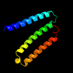
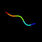
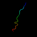
4 c3rkoE_
100.0
31
PDB header: oxidoreductaseChain: E: PDB Molecule: nadh-quinone oxidoreductase subunit a;PDBTitle: crystal structure of the membrane domain of respiratory complex i from2 e. coli at 3.0 angstrom resolution
5 c3rkoA_
100.0
31
PDB header: oxidoreductaseChain: A: PDB Molecule: nadh-quinone oxidoreductase subunit a;PDBTitle: crystal structure of the membrane domain of respiratory complex i from2 e. coli at 3.0 angstrom resolution
6 c4he8B_
100.0
41
PDB header: oxidoreductaseChain: B: PDB Molecule: nadh-quinone oxidoreductase subunit 7;PDBTitle: crystal structure of the membrane domain of respiratory complex i from2 thermus thermophilus
7 c6g72A_
99.9
27
PDB header: oxidoreductaseChain: A: PDB Molecule: nadh-ubiquinone oxidoreductase chain 3;PDBTitle: mouse mitochondrial complex i in the deactive state
8 c2ml7A_
35.2
43
PDB header: unknown functionChain: A: PDB Molecule: specific abundant protein 3;PDBTitle: ginsentides: characterization, structure and application of a new2 class of highly stable cystine knot peptides in ginseng
9 d1jwya1
26.6
31
Fold: SH3-like barrelSuperfamily: Myosin S1 fragment, N-terminal domainFamily: Myosin S1 fragment, N-terminal domain
10 d3bz7a1
24.9
31
Fold: SH3-like barrelSuperfamily: Myosin S1 fragment, N-terminal domainFamily: Myosin S1 fragment, N-terminal domain
11 c3fy6A_
21.3
33
PDB header: structural genomics, unknown functionChain: A: PDB Molecule: integron cassette protein;PDBTitle: structure from the mobile metagenome of v. cholerae. integron cassette2 protein vch_cass3
12 d2csha2
18.2
60
Fold: beta-beta-alpha zinc fingersSuperfamily: beta-beta-alpha zinc fingersFamily: Classic zinc finger, C2H2
13 d1k32a1
15.3
29
Fold: PDZ domain-likeSuperfamily: PDZ domain-likeFamily: Tail specific protease PDZ domain
14 d2adra2
14.8
50
Fold: beta-beta-alpha zinc fingersSuperfamily: beta-beta-alpha zinc fingersFamily: Classic zinc finger, C2H2
15 c2elsA_
9.3
27
PDB header: transcriptionChain: A: PDB Molecule: zinc finger protein 406;PDBTitle: solution structure of the 2nd c2h2 zinc finger of human2 zinc finger protein 406
16 c4oudA_
8.8
31
PDB header: ligaseChain: A: PDB Molecule: tyrosyl-trna synthetase;PDBTitle: engineered tyrosyl-trna synthetase with the nonstandard amino acid l-2 4,4-biphenylalanine
17 c2k9pA_
8.6
15
PDB header: membrane proteinChain: A: PDB Molecule: pheromone alpha factor receptor;PDBTitle: structure of tm1_tm2 in lppg micelles
18 c6cfwI_
8.6
15
PDB header: membrane proteinChain: I: PDB Molecule: mbh subunit;PDBTitle: cryoem structure of a respiratory membrane-bound hydrogenase
19 c1zrtD_
8.2
11
PDB header: oxidoreductase/metal transportChain: D: PDB Molecule: cytochrome c1;PDBTitle: rhodobacter capsulatus cytochrome bc1 complex with2 stigmatellin bound
20 c2ytaA_
8.0
33
PDB header: metal binding proteinChain: A: PDB Molecule: zinc finger protein 32;PDBTitle: solution structure of c2h2 type zinc finger domain 3 in2 zinc finger protein 32
21 d2j7ja2
not modelled
7.6
67
Fold: beta-beta-alpha zinc fingersSuperfamily: beta-beta-alpha zinc fingersFamily: Classic zinc finger, C2H2
22 c4oudB_
not modelled
7.5
38
PDB header: ligaseChain: B: PDB Molecule: tyrosyl-trna synthetase;PDBTitle: engineered tyrosyl-trna synthetase with the nonstandard amino acid l-2 4,4-biphenylalanine
23 c5oliA_
not modelled
7.1
6
PDB header: protein bindingChain: A: PDB Molecule: putative transferase caf17, mitochondrial;PDBTitle: crystal structure of human iba57
24 c2dmdA_
not modelled
6.6
8
PDB header: transcriptionChain: A: PDB Molecule: zinc finger protein 64, isoforms 1 and 2;PDBTitle: solution structure of the n-terminal c2h2 type zinc-binding2 domain of the zinc finger protein 64, isoforms 1 and 2
25 c2fynH_
not modelled
6.6
16
PDB header: oxidoreductaseChain: H: PDB Molecule: cytochrome c1;PDBTitle: crystal structure analysis of the double mutant rhodobacter2 sphaeroides bc1 complex
26 c3ssbC_
not modelled
6.2
100
PDB header: hydrolase/hydrolase inhibitorChain: C: PDB Molecule: inducible metalloproteinase inhibitor protein;PDBTitle: structure of insect metalloproteinase inhibitor in complex with2 thermolysin
27 c2wsc1_
not modelled
5.9
15
PDB header: photosynthesisChain: 1: PDB Molecule: at3g54890;PDBTitle: improved model of plant photosystem i
28 c2axqA_
not modelled
5.9
50
PDB header: oxidoreductaseChain: A: PDB Molecule: saccharopine dehydrogenase;PDBTitle: apo histidine-tagged saccharopine dehydrogenase (l-glu2 forming) from saccharomyces cerevisiae


