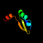
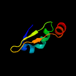
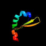
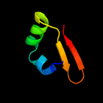
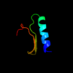
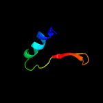
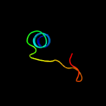
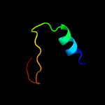
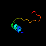
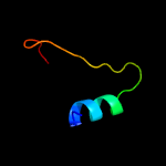
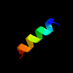
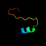
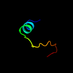
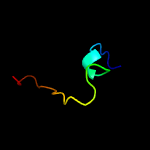
1 c2odkD_
99.4
20
PDB header: structural genomics, unknown functionChain: D: PDB Molecule: hypothetical protein;PDBTitle: putative prevent-host-death protein from nitrosomonas europaea
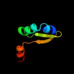
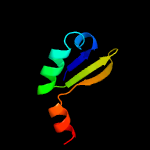
2 d2odka1
99.3
20
Fold: YefM-likeSuperfamily: YefM-likeFamily: YefM-like
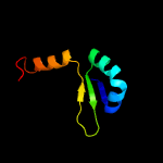
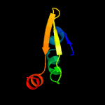
3 c3hryA_
99.2
36
PDB header: antitoxinChain: A: PDB Molecule: prevent host death protein;PDBTitle: crystal structure of phd in a trigonal space group and partially2 disordered
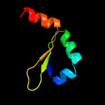
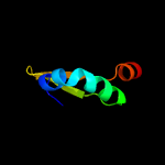
4 c3hs2H_
99.1
39
PDB header: antitoxinChain: H: PDB Molecule: prevent host death protein;PDBTitle: crystal structure of phd truncated to residue 57 in an orthorhombic2 space group
5 d2a6qb1
98.7
23
Fold: YefM-likeSuperfamily: YefM-likeFamily: YefM-like
6 c3g5oA_
98.6
20
PDB header: toxin/antitoxinChain: A: PDB Molecule: uncharacterized protein rv2865;PDBTitle: the crystal structure of the toxin-antitoxin complex relbe2 (rv2865-2 2866) from mycobacterium tuberculosis
7 d2a6qa1
98.4
23
Fold: YefM-likeSuperfamily: YefM-likeFamily: YefM-like
8 c3oeiB_
97.7
24
PDB header: toxin, protein bindingChain: B: PDB Molecule: relj (antitoxin rv3357);PDBTitle: crystal structure of mycobacterium tuberculosis reljk (rv3357-rv3358-2 relbe3)
9 c3d55A_
97.6
24
PDB header: toxin inhibitorChain: A: PDB Molecule: uncharacterized protein rv3357/mt3465;PDBTitle: crystal structure of m. tuberculosis yefm antitoxin
10 c3k6qB_
86.7
11
PDB header: ligand binding proteinChain: B: PDB Molecule: putative ligand binding protein;PDBTitle: crystal structure of an antitoxin part of a putative toxin/antitoxin2 system (swol_0700) from syntrophomonas wolfei subsp. wolfei at 1.80 a3 resolution
11 c1skoA_
62.3
20
PDB header: signaling proteinChain: A: PDB Molecule: mitogen-activated protein kinase kinase 1PDBTitle: mp1-p14 complex
12 d3cpta1
56.3
26
Fold: Profilin-likeSuperfamily: Roadblock/LC7 domainFamily: Roadblock/LC7 domain
13 d2ns0a1
33.3
29
Fold: DNA/RNA-binding 3-helical bundleSuperfamily: "Winged helix" DNA-binding domainFamily: RHA1 ro06458-like
14 d1ogda_
19.4
30
Fold: RbsD-likeSuperfamily: RbsD-likeFamily: RbsD-like
15 c2wcvI_
18.9
19
PDB header: isomeraseChain: I: PDB Molecule: l-fucose mutarotase;PDBTitle: crystal structure of bacterial fucu
16 c3e7nB_
16.8
26
PDB header: transport proteinChain: B: PDB Molecule: d-ribose high-affinity transport system;PDBTitle: crystal structure of d-ribose high-affinity transport system from2 salmonella typhimurium lt2
17 c1gk7A_
16.5
19
PDB header: vimentinChain: A: PDB Molecule: vimentin;PDBTitle: human vimentin coil 1a fragment (1a)
18 c3p13B_
16.1
22
PDB header: isomeraseChain: B: PDB Molecule: d-ribose pyranase;PDBTitle: complex structure of d-ribose pyranase sa240 with d-ribose
19 c4a34L_
14.9
19
PDB header: isomeraseChain: L: PDB Molecule: rbsd/fucu transport protein family protein;PDBTitle: crystal structure of the fucose mutarotase in complex with2 l-fucose from streptococcus pneumoniae
20 c3mvkA_
14.7
19
PDB header: isomeraseChain: A: PDB Molecule: protein fucu;PDBTitle: the crystal structure of fucu from bifidobacterium longum to 1.65a
21 c2wcuB_
not modelled
14.6
27
PDB header: isomeraseChain: B: PDB Molecule: protein fucu homolog;PDBTitle: crystal structure of mammalian fucu
22 d2ob5a1
not modelled
12.8
31
Fold: RbsD-likeSuperfamily: RbsD-likeFamily: RbsD-like
23 d1bifa2
not modelled
12.2
18
Fold: Phosphoglycerate mutase-likeSuperfamily: Phosphoglycerate mutase-likeFamily: 6-phosphofructo-2-kinase/fructose-2,6-bisphosphatase, phosphatase domain
24 c3s4rB_
not modelled
12.2
25
PDB header: structural proteinChain: B: PDB Molecule: vimentin;PDBTitle: crystal structure of vimentin coil1a/1b fragment with a stabilizing2 mutation
25 c2vm2C_
not modelled
11.6
17
PDB header: oxidoreductaseChain: C: PDB Molecule: thioredoxin h isoform 1.;PDBTitle: crystal structure of barley thioredoxin h isoform 1 crystallized using2 peg as precipitant
26 d1k6ma2
not modelled
11.5
18
Fold: Phosphoglycerate mutase-likeSuperfamily: Phosphoglycerate mutase-likeFamily: 6-phosphofructo-2-kinase/fructose-2,6-bisphosphatase, phosphatase domain
27 d1tipa_
not modelled
11.1
18
Fold: Phosphoglycerate mutase-likeSuperfamily: Phosphoglycerate mutase-likeFamily: 6-phosphofructo-2-kinase/fructose-2,6-bisphosphatase, phosphatase domain
28 d1y8xb1
not modelled
10.4
21
Fold: Activating enzymes of the ubiquitin-like proteinsSuperfamily: Activating enzymes of the ubiquitin-like proteinsFamily: Ubiquitin activating enzymes (UBA)
29 c5zf2A_
not modelled
10.1
14
PDB header: oxidoreductaseChain: A: PDB Molecule: thioredoxin (h-type,trx-h);PDBTitle: crystal structure of trxlp from edwardsiella tarda eib202
30 c4heoA_
not modelled
10.1
35
PDB header: viral proteinChain: A: PDB Molecule: phosphoprotein;PDBTitle: hendra virus phosphoprotein c terminal domain
31 d2hq7a1
not modelled
9.5
13
Fold: Split barrel-likeSuperfamily: FMN-binding split barrelFamily: PNP-oxidase like
32 c2wtoB_
not modelled
9.3
33
PDB header: metal binding proteinChain: B: PDB Molecule: orf131 protein;PDBTitle: crystal structure of apo-form czce from c. metallidurans ch34
33 d1x6va1
not modelled
8.9
27
Fold: PUA domain-likeSuperfamily: PUA domain-likeFamily: ATP sulfurylase N-terminal domain
34 c3jvoA_
not modelled
8.2
6
PDB header: viral proteinChain: A: PDB Molecule: gp6;PDBTitle: crystal structure of bacteriophage hk97 gp6
35 c2qsiB_
not modelled
8.0
9
PDB header: structural genomics, unknown functionChain: B: PDB Molecule: putative hydrogenase expression/formation protein hupg;PDBTitle: crystal structure of putative hydrogenase expression/formation protein2 hupg from rhodopseudomonas palustris cga009
36 c3iprC_
not modelled
7.7
19
PDB header: transferaseChain: C: PDB Molecule: pts system, iia component;PDBTitle: crystal structure of the enterococcus faecalis gluconate2 specific eiia phosphotransferase system component
37 c2pptA_
not modelled
7.5
10
PDB header: oxidoreductaseChain: A: PDB Molecule: thioredoxin-2;PDBTitle: crystal structure of thioredoxin-2
38 c5lddA_
not modelled
7.5
20
PDB header: protein transportChain: A: PDB Molecule: mon1;PDBTitle: crystal structure of the heterodimeric gef mon1-ccz1 in complex with2 ypt7
39 c4ewvB_
not modelled
6.8
30
PDB header: ligaseChain: B: PDB Molecule: 4-substituted benzoates-glutamate ligase gh3.12;PDBTitle: crystal structure of gh3.12 in complex with ampcpp
40 c3v62F_
not modelled
6.3
50
PDB header: protein binding/dna binding proteinChain: F: PDB Molecule: atp-dependent dna helicase srs2;PDBTitle: structure of the s. cerevisiae srs2 c-terminal domain in complex with2 pcna conjugated to sumo on lysine 164
41 c3v62C_
not modelled
6.3
50
PDB header: protein binding/dna binding proteinChain: C: PDB Molecule: atp-dependent dna helicase srs2;PDBTitle: structure of the s. cerevisiae srs2 c-terminal domain in complex with2 pcna conjugated to sumo on lysine 164
42 c4jj0B_
not modelled
5.7
17
PDB header: electron transportChain: B: PDB Molecule: mamp;PDBTitle: crystal structure of mamp
43 c4x3iA_
not modelled
5.7
30
PDB header: signaling proteinChain: A: PDB Molecule: activity-regulated cytoskeleton-associated protein;PDBTitle: the crystal structure of arc n-lobe complexed with camk2a fragment


