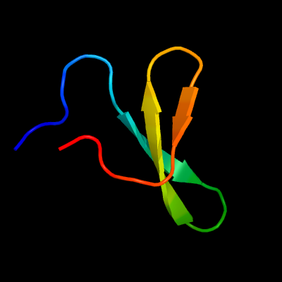
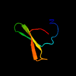
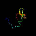
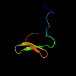
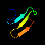
1 c4rohA_
72.3
32
PDB header: ligaseChain: A: PDB Molecule: e3 ubiquitin-protein ligase itchy homolog;PDBTitle: crystal structure of tandem ww domains of itch in complex with txnip2 peptide
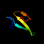
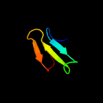
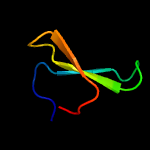
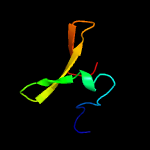
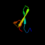
2 d2jmfa1
71.7
33
Fold: WW domain-likeSuperfamily: WW domainFamily: WW domain
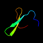
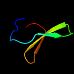
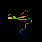
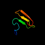
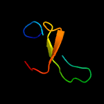
3 c3l4hA_
66.1
26
PDB header: protein bindingChain: A: PDB Molecule: e3 ubiquitin-protein ligase hecw1;PDBTitle: helical box domain and second ww domain of the human e3 ubiquitin-2 protein ligase hecw1
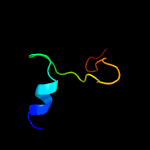
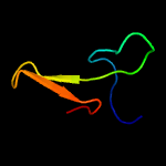
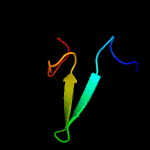
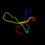
4 c1yiuA_
64.3
33
PDB header: ligaseChain: A: PDB Molecule: itchy e3 ubiquitin protein ligase;PDBTitle: itch e3 ubiquitin ligase ww3 domain
5 c2lawA_
60.2
33
PDB header: signaling protein/transcriptionChain: A: PDB Molecule: yorkie homolog;PDBTitle: structure of the second ww domain from human yap in complex with a2 human smad1 derived peptide
6 c2kykA_
59.3
33
PDB header: ligaseChain: A: PDB Molecule: e3 ubiquitin-protein ligase itchy homolog;PDBTitle: the sandwich region between two lmp2a py motif regulates the2 interaction between aip4ww2domain and py motif
7 c2ysdA_
54.8
21
PDB header: protein bindingChain: A: PDB Molecule: membrane-associated guanylate kinase, ww and pdzPDBTitle: solution structure of the first ww domain from the human2 membrane-associated guanylate kinase, ww and pdz domain-3 containing protein 1. magi-1
8 c2l4jA_
54.0
26
PDB header: transcriptionChain: A: PDB Molecule: yes-associated protein 2 (yap2);PDBTitle: yap ww2
9 c1ymzA_
52.8
31
PDB header: unknown functionChain: A: PDB Molecule: cc45;PDBTitle: cc45, an artificial ww domain designed using statistical2 coupling analysis
10 c2ez5W_
52.7
18
PDB header: signalling protein,ligaseChain: W: PDB Molecule: e3 ubiquitin-protein ligase nedd4;PDBTitle: solution structure of the dnedd4 ww3* domain- comm lpsy2 peptide complex
11 c2djyA_
51.9
30
PDB header: ligase/signaling proteinChain: A: PDB Molecule: smad ubiquitination regulatory factor 2;PDBTitle: solution structure of smurf2 ww3 domain-smad7 py peptide2 complex
12 d1i5hw_
50.0
24
Fold: WW domain-likeSuperfamily: WW domainFamily: WW domain
13 c2ysbA_
49.2
26
PDB header: protein bindingChain: A: PDB Molecule: salvador homolog 1 protein;PDBTitle: solution structure of the first ww domain from the mouse2 salvador homolog 1 protein (sav1)
14 d1tk7a1
48.0
22
Fold: WW domain-likeSuperfamily: WW domainFamily: WW domain
15 c2jmfA_
45.7
33
PDB header: ligase/signaling proteinChain: A: PDB Molecule: e3 ubiquitin-protein ligase suppressor of deltex;PDBTitle: solution structure of the su(dx) ww4- notch py peptide2 complex
16 c1wr7A_
44.7
27
PDB header: ligaseChain: A: PDB Molecule: nedd4-2;PDBTitle: solution structure of the third ww domain of nedd4-2
17 d1tk7a2
44.5
33
Fold: WW domain-likeSuperfamily: WW domainFamily: WW domain
18 c5ydyA_
43.7
30
PDB header: signaling proteinChain: A: PDB Molecule: ww2 domain and ppxy motif complex;PDBTitle: nmr structure of yap1-2 ww2 domain with lats1 ppxy motif complex
19 c2mdjA_
42.9
26
PDB header: transferaseChain: A: PDB Molecule: histone-lysine n-methyltransferase setd2;PDBTitle: solution structure of ww domain with polyproline stretch (pp2ww) of2 hypb
20 d1f8ab1
41.8
25
Fold: WW domain-likeSuperfamily: WW domainFamily: WW domain
21 c2n8tA_
not modelled
41.2
25
PDB header: ligase/peptideChain: A: PDB Molecule: e3 ubiquitin-protein ligase nedd4;PDBTitle: solution structure of the rnedd4 ww2 domain-cx43ct peptide complex by2 nmr
22 c1wmvA_
not modelled
40.9
27
PDB header: oxidoreductase, apoptosisChain: A: PDB Molecule: ww domain containing oxidoreductase;PDBTitle: solution structure of the second ww domain of wwox
23 c2dmvA_
not modelled
40.2
24
PDB header: ligaseChain: A: PDB Molecule: itchy homolog e3 ubiquitin protein ligase;PDBTitle: solution structure of the second ww domain of itchy homolog2 e3 ubiquitin protein ligase (itch)
24 c1wr4A_
not modelled
40.0
31
PDB header: ligaseChain: A: PDB Molecule: ubiquitin-protein ligase nedd4-2;PDBTitle: solution structure of the second ww domain of nedd4-2
25 c2e3uA_
not modelled
39.6
27
PDB header: rna binding proteinChain: A: PDB Molecule: hypothetical protein ph1566;PDBTitle: crystal structure analysis of dim2p from pyrococcus horikoshii ot3
26 c1e0mA_
not modelled
39.5
30
PDB header: de novo proteinChain: A: PDB Molecule: wwprototype;PDBTitle: prototype ww domain
27 c2ysgA_
not modelled
37.7
24
PDB header: protein bindingChain: A: PDB Molecule: syntaxin-binding protein 4;PDBTitle: solution structure of the ww domain from the human syntaxin-2 binding protein 4
28 c1tuaA_
not modelled
37.1
25
PDB header: structural genomics, unknown functionChain: A: PDB Molecule: hypothetical protein ape0754;PDBTitle: 1.5 a crystal structure of a protein of unknown function2 ape0754 from aeropyrum pernix
29 c2dwvB_
not modelled
33.8
17
PDB header: protein bindingChain: B: PDB Molecule: salvador homolog 1 protein;PDBTitle: solution structure of the second ww domain from mouse2 salvador homolog 1 protein (mww45)
30 c2ysfA_
not modelled
29.2
18
PDB header: protein bindingChain: A: PDB Molecule: e3 ubiquitin-protein ligase itchy homolog;PDBTitle: solution structure of the fourth ww domain from the human2 e3 ubiquitin-protein ligase itchy homolog, itch
31 d1k9ra_
not modelled
29.0
35
Fold: WW domain-likeSuperfamily: WW domainFamily: WW domain
32 c2kq0A_
not modelled
27.5
25
PDB header: ligaseChain: A: PDB Molecule: e3 ubiquitin-protein ligase nedd4;PDBTitle: human nedd4 3rd ww domain complex with ebola zaire virus matrix2 protein vp40 derived peptide ilptappeymea
33 d1tuaa2
not modelled
26.6
25
Fold: Eukaryotic type KH-domain (KH-domain type I)Superfamily: Eukaryotic type KH-domain (KH-domain type I)Family: Eukaryotic type KH-domain (KH-domain type I)
34 c2c36B_
not modelled
26.4
22
PDB header: viral proteinChain: B: PDB Molecule: glycoprotein d hsv-1;PDBTitle: structure of unliganded hsv gd reveals a mechanism for receptor-2 mediated activation of virus entry
35 c1tk7A_
not modelled
26.1
27
PDB header: signaling proteinChain: A: PDB Molecule: cg4244-pb;PDBTitle: nmr structure of ww domains (ww3-4) from suppressor of2 deltex
36 c4b8tA_
not modelled
25.4
22
PDB header: transcription/rnaChain: A: PDB Molecule: kh-type splicing regulatory protein;PDBTitle: rna binding protein solution structure of the third kh2 domain of ksrp in complex with the g-rich target sequence.
37 c6g18x_
not modelled
24.3
17
PDB header: ribosomeChain: X: PDB Molecule: 40s ribosomal protein s23;PDBTitle: cryo-em structure of a late human pre-40s ribosomal subunit - state c
38 c5ydxA_
not modelled
24.1
38
PDB header: signaling proteinChain: A: PDB Molecule: ww domain with ppxy motif;PDBTitle: nmr structure of yap1-2 ww1 domain with lats1 ppxy motif complex
39 d1nmva1
not modelled
23.5
24
Fold: WW domain-likeSuperfamily: WW domainFamily: WW domain
40 c2dl0A_
not modelled
23.3
32
PDB header: signaling proteinChain: A: PDB Molecule: sam and sh3 domain-containing protein 1;PDBTitle: solution structure of the sam-domain of the sam and sh32 domain containing protein 1
41 d1dt4a_
not modelled
23.0
28
Fold: Eukaryotic type KH-domain (KH-domain type I)Superfamily: Eukaryotic type KH-domain (KH-domain type I)Family: Eukaryotic type KH-domain (KH-domain type I)
42 d2itka1
not modelled
21.6
25
Fold: WW domain-likeSuperfamily: WW domainFamily: WW domain
43 c2yshA_
not modelled
20.8
29
PDB header: protein bindingChain: A: PDB Molecule: growth-arrest-specific protein 7;PDBTitle: solution structure of the ww domain from the human growth-2 arrest-specific protein 7, gas-7
44 c2yscA_
not modelled
19.9
37
PDB header: protein bindingChain: A: PDB Molecule: amyloid beta a4 precursor protein-binding familyPDBTitle: solution structure of the ww domain from the human amyloid2 beta a4 precursor protein-binding family b member 3, apbb3
45 d2f21a1
not modelled
19.8
29
Fold: WW domain-likeSuperfamily: WW domainFamily: WW domain
46 c2kxqA_
not modelled
19.3
18
PDB header: protein bindingChain: A: PDB Molecule: e3 ubiquitin-protein ligase smurf2;PDBTitle: solution structure of smurf2 ww2 and ww3 bound to smad7 py motif2 containing peptide
47 c2ysiA_
not modelled
16.6
17
PDB header: protein bindingChain: A: PDB Molecule: transcription elongation regulator 1;PDBTitle: solution structure of the first ww domain from the mouse2 transcription elongation regulator 1, transcription factor3 ca150
48 d1ec6a_
not modelled
16.3
31
Fold: Eukaryotic type KH-domain (KH-domain type I)Superfamily: Eukaryotic type KH-domain (KH-domain type I)Family: Eukaryotic type KH-domain (KH-domain type I)
49 d2ho2a1
not modelled
15.6
43
Fold: WW domain-likeSuperfamily: WW domainFamily: WW domain
50 d1pina1
not modelled
15.1
28
Fold: WW domain-likeSuperfamily: WW domainFamily: WW domain
51 c2lb0A_
not modelled
15.0
21
PDB header: signaling protein/transcriptionChain: A: PDB Molecule: e3 ubiquitin-protein ligase smurf1;PDBTitle: structure of the first ww domain of human smurf1 in complex with a di-2 phosphorylated human smad1 derived peptide
52 d2dsya1
not modelled
14.7
33
Fold: TTHA1013/TTHA0281-likeSuperfamily: TTHA1013/TTHA0281-likeFamily: TTHA0281-like
53 c6emlp_
not modelled
14.2
17
PDB header: ribosomeChain: P: PDB Molecule: 40s ribosomal protein s0-a;PDBTitle: cryo-em structure of a late pre-40s ribosomal subunit from2 saccharomyces cerevisiae
54 c2zajA_
not modelled
13.7
19
PDB header: protein bindingChain: A: PDB Molecule: membrane-associated guanylate kinase, ww and pdzPDBTitle: solution structure of the short-isoform of the second ww2 domain from the human membrane-associated guanylate kinase,3 ww and pdz domain-containing protein 1 (magi-1)
55 d1i8gb_
not modelled
13.4
29
Fold: WW domain-likeSuperfamily: WW domainFamily: WW domain
56 d1dtja_
not modelled
13.1
26
Fold: Eukaryotic type KH-domain (KH-domain type I)Superfamily: Eukaryotic type KH-domain (KH-domain type I)Family: Eukaryotic type KH-domain (KH-domain type I)
57 c2lazA_
not modelled
12.9
21
PDB header: signaling protein/transcriptionChain: A: PDB Molecule: e3 ubiquitin-protein ligase smurf1;PDBTitle: structure of the first ww domain of human smurf1 in complex with a2 mono-phosphorylated human smad1 derived peptide
58 d1x4ma1
not modelled
11.8
12
Fold: Eukaryotic type KH-domain (KH-domain type I)Superfamily: Eukaryotic type KH-domain (KH-domain type I)Family: Eukaryotic type KH-domain (KH-domain type I)
59 c5u3kC_
not modelled
11.1
22
PDB header: immune system/viral proteinChain: C: PDB Molecule: gp41 mper peptide;PDBTitle: crystal structure of dh511.2 fab in complex with hiv-1 gp41 mper 662-2 683 peptide
60 d2ysca1
not modelled
10.8
41
Fold: WW domain-likeSuperfamily: WW domainFamily: WW domain
61 c5u3kP_
not modelled
10.6
22
PDB header: immune system/viral proteinChain: P: PDB Molecule: gp41 mper peptide;PDBTitle: crystal structure of dh511.2 fab in complex with hiv-1 gp41 mper 662-2 683 peptide
62 c1loiA_
not modelled
10.4
100
PDB header: hydrolaseChain: A: PDB Molecule: cyclic 3',5'-amp specific phosphodiesterase rd1;PDBTitle: n-terminal splice region of rat c-amp phosphodiesterase,2 nmr, 7 structures
63 c2w0cS_
not modelled
10.3
29
PDB header: virusChain: S: PDB Molecule: protein p3;PDBTitle: x-ray structure of the entire lipid-containing bacteriophage pm2
64 c6htnE_
not modelled
10.3
23
PDB header: sugar binding proteinChain: E: PDB Molecule: fucose-binding lectin;PDBTitle: structure of a fucose lectin from kordia zhangzhouensis in complex2 with methyl-fucoside
65 d1xmeb2
not modelled
10.2
35
Fold: Transmembrane helix hairpinSuperfamily: Cytochrome c oxidase subunit II-like, transmembrane regionFamily: Cytochrome c oxidase subunit II-like, transmembrane region
66 c2hh2A_
not modelled
9.7
25
PDB header: rna binding proteinChain: A: PDB Molecule: kh-type splicing regulatory protein;PDBTitle: solution structure of the fourth kh domain of ksrp
67 c1javA_
not modelled
9.2
22
PDB header: viral proteinChain: A: PDB Molecule: transmembrane glycoprotein (gp41);PDBTitle: average nmr solution structure of the trp-rich peptide of2 hiv gp41 bound to dpc micelles
68 c2g57A_
not modelled
8.9
67
PDB header: oncoproteinChain: A: PDB Molecule: beta-catenin;PDBTitle: structure of the phosphorylation motif of the oncogenic2 protein beta-catenin recognized by a selective monoclonal3 antibody
69 c1jauA_
not modelled
8.8
22
PDB header: viral proteinChain: A: PDB Molecule: transmembrane glycoprotein (gp41);PDBTitle: nmr solution structure of the trp-rich peptide of hiv gp412 bound to dpc micelles
70 d3bmva3
not modelled
8.7
56
Fold: Glycosyl hydrolase domainSuperfamily: Glycosyl hydrolase domainFamily: alpha-Amylases, C-terminal beta-sheet domain
71 d1smpi_
not modelled
7.9
39
Fold: Streptavidin-likeSuperfamily: beta-Barrel protease inhibitorsFamily: Metalloprotease inhibitor
72 c6mttP_
not modelled
7.8
22
PDB header: immune systemChain: P: PDB Molecule: rv217 founder virus gp41 peptide;PDBTitle: crystal structure of vrc46.01 fab in complex with gp41 peptide
73 c2dgrA_
not modelled
7.6
18
PDB header: rna binding proteinChain: A: PDB Molecule: ring finger and kh domain-containing protein 1;PDBTitle: solution structure of the second kh domain in ring finger2 and kh domain containing protein 1
74 d1lgha_
not modelled
7.4
43
Fold: Light-harvesting complex subunitsSuperfamily: Light-harvesting complex subunitsFamily: Light-harvesting complex subunits
75 c2pv6A_
not modelled
7.3
22
PDB header: viral proteinChain: A: PDB Molecule: envelope glycoprotein;PDBTitle: hiv-1 gp41 membrane proximal ectodomain region peptide in2 dpc micelle
76 c6acvA_
not modelled
7.1
15
PDB header: dna binding proteinChain: A: PDB Molecule: methyl-cpg-binding domain-containing protein 11;PDBTitle: the solution nmr structure of mbd domain
77 c4ce41_
not modelled
6.9
50
PDB header: ribosomeChain: 1: PDB Molecule: mrpl28;PDBTitle: 39s large subunit of the porcine mitochondrial ribosome
78 c5wvmA_
not modelled
6.7
35
PDB header: sugar binding proteinChain: A: PDB Molecule: maltose-binding periplasmic protein,two-component systemPDBTitle: crystal structure of baes cocrystallized with 2 mm indole
79 c4pqkA_
not modelled
6.7
55
PDB header: dna binding proteinChain: A: PDB Molecule: maltose abc transporter periplasmic protein, truncatedPDBTitle: c-terminal domain of dna binding protein
80 c4v3aC_
not modelled
6.5
70
PDB header: transport proteinChain: C: PDB Molecule: pleurotolysin b;PDBTitle: membrane bound pleurotolysin prepore (tmh1 lock) trapped with2 engineered disulphide cross-link
81 c3j20W_
not modelled
6.4
38
PDB header: ribosomeChain: W: PDB Molecule: 30s ribosomal protein s27e;PDBTitle: promiscuous behavior of proteins in archaeal ribosomes revealed by2 cryo-em: implications for evolution of eukaryotic ribosomes (30s3 ribosomal subunit)
82 d1j4wa2
not modelled
6.4
22
Fold: Eukaryotic type KH-domain (KH-domain type I)Superfamily: Eukaryotic type KH-domain (KH-domain type I)Family: Eukaryotic type KH-domain (KH-domain type I)
83 c2me1A_
not modelled
6.4
22
PDB header: membrane proteinChain: A: PDB Molecule: gp41;PDBTitle: hiv-1 gp41 clade b double alanine mutant membrane proximal external2 region peptide in dpc micelle
84 c4ol4A_
not modelled
6.0
13
PDB header: lipid binding proteinChain: A: PDB Molecule: proline-rich 28 kda antigen;PDBTitle: crystal structure of secreted proline rich antigen mtc28 (rv0040c)2 from mycobacterium tuberculosis
85 c3cglF_
not modelled
5.9
56
PDB header: fluorescent proteinChain: F: PDB Molecule: gfp-like fluorescent chromoprotein dsfp483;PDBTitle: crystal structure and raman studies of dsfp483, a cyan fluorescent2 protein from discosoma striata
86 c2mnsA_
not modelled
5.8
36
PDB header: membrane proteinChain: A: PDB Molecule: membrane fusion protein p15;PDBTitle: solution nmr structure of the reovirus p15 fusion-associated small2 transmembrane (fast) protein fusion-inducing lipid packing sensor3 (flips) motif in dodecyl phosphocholine (dpc) micelles
87 d2ctla1
not modelled
5.7
29
Fold: Eukaryotic type KH-domain (KH-domain type I)Superfamily: Eukaryotic type KH-domain (KH-domain type I)Family: Eukaryotic type KH-domain (KH-domain type I)
88 d1wvna1
not modelled
5.7
18
Fold: Eukaryotic type KH-domain (KH-domain type I)Superfamily: Eukaryotic type KH-domain (KH-domain type I)Family: Eukaryotic type KH-domain (KH-domain type I)
89 c4p1nA_
not modelled
5.6
36
PDB header: protein transportChain: A: PDB Molecule: atg1 tmit;PDBTitle: crystal structure of atg1-atg13 complex
90 c2jniA_
not modelled
5.6
83
PDB header: antimicrobial proteinChain: A: PDB Molecule: arenicin-2;PDBTitle: spatial structure of antimicrobial peptide arenicin-2 in2 aqueous solution
91 c2l8xB_
not modelled
5.6
83
PDB header: antimicrobial proteinChain: B: PDB Molecule: arenicin-2;PDBTitle: spatial structure of antimicrobial peptide arenicin-2 dimer in dpc2 micelles
92 c2jsbA_
not modelled
5.6
83
PDB header: antimicrobial proteinChain: A: PDB Molecule: arenicin-1;PDBTitle: solution structure of arenicin-1
93 c2y5pB_
not modelled
5.5
22
PDB header: protein bindingChain: B: PDB Molecule: internalin b;PDBTitle: b-repeat of listeria monocytogenes inlb (internalin b)
94 c3uiyA_
not modelled
5.4
50
PDB header: structural proteinChain: A: PDB Molecule: chimera protein of sefd and sefa;PDBTitle: crystal structure of sefd_dsca in h2o
95 c2l8xA_
not modelled
5.4
83
PDB header: antimicrobial proteinChain: A: PDB Molecule: arenicin-2;PDBTitle: spatial structure of antimicrobial peptide arenicin-2 dimer in dpc2 micelles
96 c2jzxA_
not modelled
5.3
23
PDB header: rna binding proteinChain: A: PDB Molecule: poly(rc)-binding protein 2;PDBTitle: pcbp2 kh1-kh2 domains
97 c3vxvA_
not modelled
5.1
67
PDB header: hydrolase/dnaChain: A: PDB Molecule: methyl-cpg-binding domain protein 4;PDBTitle: crystal structure of methyl cpg binding domain of mbd4 in complex with2 the 5mcg/tg sequence