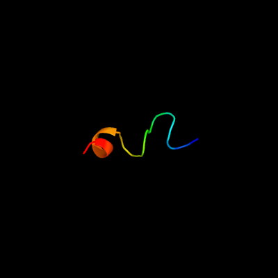
| 1 |
|
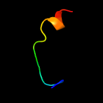
PDB 1z9i chain A
Region: 18 - 36
Aligned: 19
Modelled: 19
Confidence: 10.5%
Identity: 32%
PDB header:transferase
Chain: A: PDB Molecule:epidermal growth factor receptor;
PDBTitle: a structural model for the membrane-bound form of the2 juxtamembrane domain of the epidermal growth factor3 receptor
Phyre2
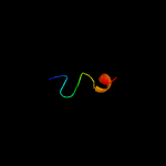
| 2 |
|
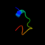
PDB 2l1q chain A
Region: 4 - 18
Aligned: 14
Modelled: 15
Confidence: 8.2%
Identity: 43%
PDB header:antimicrobial protein
Chain: A: PDB Molecule:liver-expressed antimicrobial peptide 2;
PDBTitle: solution structure of human liver expressed antimicrobial peptide 2
Phyre2
| 3 |
|
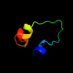
PDB 3udi chain A
Region: 9 - 23
Aligned: 15
Modelled: 15
Confidence: 7.8%
Identity: 47%
PDB header:penicillin-binding protein/antibiotic
Chain: A: PDB Molecule:penicillin-binding protein 1a;
PDBTitle: crystal structure of acinetobacter baumannii pbp1a in complex with2 penicillin g
Phyre2
| 4 |
|
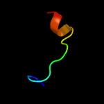
PDB 3pbq chain A
Region: 9 - 23
Aligned: 15
Modelled: 15
Confidence: 6.2%
Identity: 40%
PDB header:hydrolase/antibiotic
Chain: A: PDB Molecule:penicillin-binding protein 3;
PDBTitle: crystal structure of pbp3 complexed with imipenem
Phyre2
| 5 |
|
PDB 2xa6 chain B
Region: 36 - 50
Aligned: 15
Modelled: 15
Confidence: 5.9%
Identity: 47%
PDB header:transcription
Chain: B: PDB Molecule:kh domain-containing\,rna-binding\,signal transduction-
PDBTitle: structural basis for homodimerization of the src-associated during2 mitosis, 68 kd protein (sam68) qua1 domain
Phyre2
| 6 |
|
PDB 1wrj chain A
Region: 6 - 37
Aligned: 31
Modelled: 32
Confidence: 5.6%
Identity: 23%
PDB header:transferase
Chain: A: PDB Molecule:methylated-dna--protein-cysteine
PDBTitle: crystal structure of o6-methylguanine methyltransferase2 from sulfolobus tokodaii
Phyre2
| 7 |
|
PDB 4oon chain A
Region: 9 - 23
Aligned: 15
Modelled: 15
Confidence: 5.6%
Identity: 53%
PDB header:transferase/transferase inhibitor
Chain: A: PDB Molecule:penicillin-binding protein 1a;
PDBTitle: crystal structure of pbp1a in complex with compound 17 ((4z,8s,11e,2 14s)-5-(2-amino-1,3-thiazol-4-yl)-14-(5,6-dihydroxy-1,3-dioxo-1,3-3 dihydro-2h-isoindol-2-yl)-8-formyl-2-methyl-6-oxo-3,10-dioxa-4,7,11-4 triazatetradeca-4,11-diene-2,12,14-tricarboxylic acid)
Phyre2
| 8 |
|
PDB 2bg1 chain A domain 1
Region: 9 - 23
Aligned: 15
Modelled: 15
Confidence: 5.2%
Identity: 40%
Fold: beta-lactamase/transpeptidase-like
Superfamily: beta-lactamase/transpeptidase-like
Family: beta-Lactamase/D-ala carboxypeptidase
Phyre2