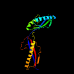
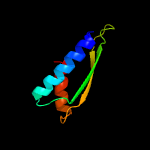
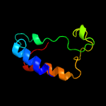
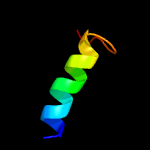
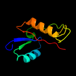
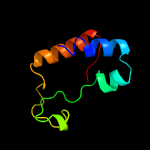
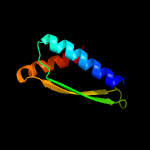
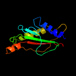
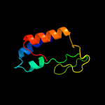
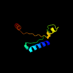
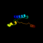
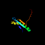
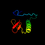
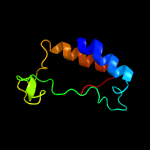
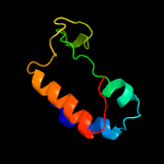
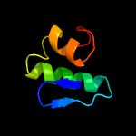
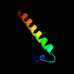
1 c4hvzA_
100.0
27
PDB header: membrane proteinChain: A: PDB Molecule: 26 kda periplasmic immunogenic protein;PDBTitle: crystal structure of brucella abortus immunogenic bp26 protein
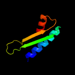
2 d1nyed_
50.2
17
Fold: OsmC-likeSuperfamily: OsmC-likeFamily: Ohr/OsmC resistance proteins
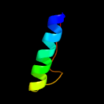
3 c3k1dA_
27.7
17
PDB header: transferaseChain: A: PDB Molecule: 1,4-alpha-glucan-branching enzyme;PDBTitle: crystal structure of glycogen branching enzyme synonym: 1,4-alpha-d-2 glucan:1,4-alpha-d-glucan 6-glucosyl-transferase from mycobacterium3 tuberculosis h37rv
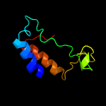
4 c2kxhB_
21.3
48
PDB header: protein bindingChain: B: PDB Molecule: peptide of far upstream element-binding protein 1;PDBTitle: solution structure of the first two rrm domains of fir in the complex2 with fbp nbox peptide
5 d1k7ka_
20.8
15
Fold: Anticodon-binding domain-likeSuperfamily: ITPase-likeFamily: ITPase (Ham1)
6 c1m7xC_
19.3
11
PDB header: transferaseChain: C: PDB Molecule: 1,4-alpha-glucan branching enzyme;PDBTitle: the x-ray crystallographic structure of branching enzyme
7 d2onfa1
18.8
14
Fold: OsmC-likeSuperfamily: OsmC-likeFamily: Ohr/OsmC resistance proteins
8 c2lfpA_
18.7
15
PDB header: viral proteinChain: A: PDB Molecule: bacteriophage spp1 complete nucleotide sequence;PDBTitle: structure of bacteriophage spp1 gp17 protein
9 c3amlA_
18.5
18
PDB header: transferaseChain: A: PDB Molecule: os06g0726400 protein;PDBTitle: structure of the starch branching enzyme i (bei) from oryza sativa l
10 c3s0yA_
18.2
8
PDB header: motor proteinChain: A: PDB Molecule: motility protein b;PDBTitle: the crystal structure of the periplasmic domain of motb (residues 64-2 256).
11 c3s0wB_
17.1
11
PDB header: motor proteinChain: B: PDB Molecule: motility protein b;PDBTitle: the crystal structure of the periplasmic domain of helicobacter pylori2 motb (residues 78-256).
12 c2rrlA_
16.0
10
PDB header: protein transportChain: A: PDB Molecule: flagellar hook-length control protein;PDBTitle: solution structure of the c-terminal domain of the flik
13 d2c8ma1
15.9
27
Fold: Class II aaRS and biotin synthetasesSuperfamily: Class II aaRS and biotin synthetasesFamily: LplA-like
14 d1ukka_
15.7
20
Fold: OsmC-likeSuperfamily: OsmC-likeFamily: Ohr/OsmC resistance proteins
15 c2zf8A_
14.5
17
PDB header: structural proteinChain: A: PDB Molecule: component of sodium-driven polar flagellar motor;PDBTitle: crystal structure of moty
16 d1m7xa3
14.1
11
Fold: TIM beta/alpha-barrelSuperfamily: (Trans)glycosidasesFamily: Amylase, catalytic domain
17 c5gquA_
13.6
11
PDB header: transferaseChain: A: PDB Molecule: 1,4-alpha-glucan branching enzyme glgb;PDBTitle: crystal structure of branching enzyme from cyanothece sp. atcc 51142
18 c4bzyC_
13.3
14
PDB header: transferaseChain: C: PDB Molecule: 1,4-alpha-glucan-branching enzyme;PDBTitle: crystal structure of human glycogen branching enzyme (gbe1)
19 c2x48B_
12.1
25
PDB header: viral proteinChain: B: PDB Molecule: cag38821;PDBTitle: orf 55 from sulfolobus islandicus rudivirus 1
20 c5yrxA_
11.8
24
PDB header: dna binding proteinChain: A: PDB Molecule: nucleoid-associated protein rv3716c;PDBTitle: crystal structure of a hypothetical protein rv3716c from mycobacterium2 tuberculosis
21 d1u07a_
not modelled
11.4
17
Fold: TolA/TonB C-terminal domainSuperfamily: TolA/TonB C-terminal domainFamily: TonB
22 c3amkA_
not modelled
11.4
18
PDB header: transferaseChain: A: PDB Molecule: os06g0726400 protein;PDBTitle: structure of the starch branching enzyme i (bei) from oryza sativa l
23 c2dgaA_
not modelled
11.3
17
PDB header: hydrolaseChain: A: PDB Molecule: beta-glucosidase;PDBTitle: crystal structure of hexameric beta-glucosidase in wheat
24 c6d9nA_
not modelled
11.0
16
PDB header: oxidoreductaseChain: A: PDB Molecule: organic hydroperoxide resistance protein;PDBTitle: crystal structure of an organic hydroperoxide resistance protein from2 elizabethkingia anophelis with crystallant-derived thiocyanate bound
25 c2wskA_
not modelled
10.9
14
PDB header: hydrolaseChain: A: PDB Molecule: glycogen debranching enzyme;PDBTitle: crystal structure of glycogen debranching enzyme glgx from2 escherichia coli k-12
26 c3mgjA_
not modelled
10.9
14
PDB header: structural genomics, unknown functionChain: A: PDB Molecule: uncharacterized protein mj1480;PDBTitle: crystal structure of the saccharop_dh_n domain of mj1480 protein from2 methanococcus jannaschii. northeast structural genomics consortium3 target mjr83a.
27 c5wtlB_
not modelled
10.8
18
PDB header: membrane proteinChain: B: PDB Molecule: ompa family protein;PDBTitle: crystal structure of the periplasmic portion of outer membrane protein2 a (ompa) from capnocytophaga gingivalis
28 c4jhoA_
not modelled
10.6
19
PDB header: hydrolaseChain: A: PDB Molecule: beta-mannosidase/beta-glucosidase;PDBTitle: structural analysis and insights into glycon specificity of the rice2 gh1 os7bglu26 beta-d-mannosidase
29 c6mjnC_
not modelled
10.5
17
PDB header: oxidoreductaseChain: C: PDB Molecule: organic hydroperoxide resistance protein;PDBTitle: crystal structure of an organic hydroperoxide resistance protein osmc,2 predicted redox protein, regulator of sulfide bond formation from3 legionella pneumophila
30 c2o0i1_
not modelled
10.2
10
PDB header: surface active proteinChain: 1: PDB Molecule: c protein alpha-antigen;PDBTitle: crystal structure of the r185a mutant of the n-terminal domain of the2 group b streptococcus alpha c protein
31 c1mg1A_
not modelled
10.1
27
PDB header: viral proteinChain: A: PDB Molecule: protein (htlv-1 gp21 ectodomain/maltose-binding proteinPDBTitle: htlv-1 gp21 ectodomain/maltose-binding protein chimera
32 d1jjcb4
not modelled
9.8
12
Fold: Ferredoxin-likeSuperfamily: Anticodon-binding domain of PheRSFamily: Anticodon-binding domain of PheRS
33 d2opla1
not modelled
9.7
13
Fold: OsmC-likeSuperfamily: OsmC-likeFamily: Ohr/OsmC resistance proteins
34 c3cyqM_
not modelled
8.8
8
PDB header: membrane proteinChain: M: PDB Molecule: chemotaxis protein motb;PDBTitle: the crystal structure of the complex of the c-terminal domain of2 helicobacter pylori motb (residues 125-256) with n-acetylmuramic acid
35 c1zb8B_
not modelled
8.7
14
PDB header: oxidoreductaseChain: B: PDB Molecule: organic hydroperoxide resistance protein;PDBTitle: crystal structure of xylella fastidiosa organic peroxide resistance2 protein
36 c2ql8A_
not modelled
8.4
11
PDB header: oxidoreductaseChain: A: PDB Molecule: putative redox protein;PDBTitle: crystal structure of a putative redox protein (lsei_0423) from2 lactobacillus casei atcc 334 at 1.50 a resolution
37 c1lqlE_
not modelled
8.1
12
PDB header: unknown functionChain: E: PDB Molecule: osmotical inducible protein c like family;PDBTitle: crystal structure of osmc like protein from mycoplasma2 pneumoniae
38 d1lqla_
not modelled
8.1
12
Fold: OsmC-likeSuperfamily: OsmC-likeFamily: Ohr/OsmC resistance proteins
39 c6izhE_
not modelled
7.7
18
PDB header: hydrolaseChain: E: PDB Molecule: 2-aminomuconate deaminase;PDBTitle: crystal structure of deaminase amne from pseudomonas sp. ap-3
40 c2grxC_
not modelled
7.7
17
PDB header: metal transportChain: C: PDB Molecule: protein tonb;PDBTitle: crystal structure of tonb in complex with fhua, e. coli2 outer membrane receptor for ferrichrome
41 c2b99A_
not modelled
7.6
17
PDB header: transferaseChain: A: PDB Molecule: riboflavin synthase;PDBTitle: crystal structure of an archaeal pentameric riboflavin2 synthase complex with a substrate analog inhibitor
42 c4y7jE_
not modelled
7.6
17
PDB header: membrane protein,transport proteinChain: E: PDB Molecule: large conductance mechanosensitive channel protein,PDBTitle: structure of an archaeal mechanosensitive channel in expanded state
43 c1bf2A_
not modelled
7.2
17
PDB header: hydrolaseChain: A: PDB Molecule: isoamylase;PDBTitle: structure of pseudomonas isoamylase
44 d2aizp1
not modelled
7.2
20
Fold: Bacillus chorismate mutase-likeSuperfamily: OmpA-likeFamily: OmpA-like
45 c1qhoA_
not modelled
7.1
19
PDB header: hydrolaseChain: A: PDB Molecule: alpha-amylase;PDBTitle: five-domain alpha-amylase from bacillus stearothermophilus,2 maltose/acarbose complex
46 c4f7dA_
not modelled
6.8
17
PDB header: oxidoreductaseChain: A: PDB Molecule: ferredoxin--nadp reductase;PDBTitle: crystal structure of ferredoxin-nadp reductase from burkholderia2 thailandensis e264
47 c2mblA_
not modelled
6.7
12
PDB header: de novo proteinChain: A: PDB Molecule: top7 fold protein top7m13;PDBTitle: solution nmr structure of de novo designed top7 fold protein top7m13,2 northeast structural genomics consortium (nesg) target or33
48 d1e4mm_
not modelled
6.6
15
Fold: TIM beta/alpha-barrelSuperfamily: (Trans)glycosidasesFamily: Family 1 of glycosyl hydrolase
49 c1xx3A_
not modelled
6.4
17
PDB header: transport proteinChain: A: PDB Molecule: tonb protein;PDBTitle: solution structure of escherichia coli tonb-ctd
50 c3m07A_
not modelled
6.4
15
PDB header: unknown functionChain: A: PDB Molecule: putative alpha amylase;PDBTitle: 1.4 angstrom resolution crystal structure of putative alpha amylase2 from salmonella typhimurium.
51 c5zu0B_
not modelled
6.2
15
PDB header: transferaseChain: B: PDB Molecule: protein arginine methyltransferase ndufaf7 homolog,PDBTitle: proteobacterial origin of protein arginine methylation and regulation2 of complex i assembly by mida
52 c2by0A_
not modelled
6.2
15
PDB header: hydrolaseChain: A: PDB Molecule: maltooligosyltrehalose trehalohydrolase;PDBTitle: is radiation damage dependent on the dose-rate used during2 macromolecular crystallography data collection
53 d2hqsc1
not modelled
6.1
18
Fold: Bacillus chorismate mutase-likeSuperfamily: OmpA-likeFamily: OmpA-like
54 c2l26A_
not modelled
6.0
27
PDB header: membrane proteinChain: A: PDB Molecule: uncharacterized protein rv0899/mt0922;PDBTitle: rv0899 from mycobacterium tuberculosis contains two separated domains
55 c3mhyC_
not modelled
5.9
10
PDB header: signaling proteinChain: C: PDB Molecule: pii-like protein pz;PDBTitle: a new pii protein structure
56 d1qwia_
not modelled
5.6
17
Fold: OsmC-likeSuperfamily: OsmC-likeFamily: Ohr/OsmC resistance proteins
57 d1uoka2
not modelled
5.6
19
Fold: TIM beta/alpha-barrelSuperfamily: (Trans)glycosidasesFamily: Amylase, catalytic domain
58 c2x7jA_
not modelled
5.6
13
PDB header: transferaseChain: A: PDB Molecule: 2-succinyl-5-enolpyruvyl-6-hydroxy-3-cyclohexenePDBTitle: structure of the menaquinone biosynthesis protein mend from2 bacillus subtilis
59 c3bmwA_
not modelled
5.6
17
PDB header: transferaseChain: A: PDB Molecule: cyclomaltodextrin glucanotransferase;PDBTitle: cyclodextrin glycosyl transferase from thermoanerobacterium2 thermosulfurigenes em1 mutant s77p complexed with a maltoheptaose3 inhibitor
60 c1nvmB_
not modelled
5.3
21
PDB header: lyase/oxidoreductaseChain: B: PDB Molecule: acetaldehyde dehydrogenase (acylating);PDBTitle: crystal structure of a bifunctional aldolase-dehydrogenase :2 sequestering a reactive and volatile intermediate
61 d1n2fa_
not modelled
5.2
11
Fold: OsmC-likeSuperfamily: OsmC-likeFamily: Ohr/OsmC resistance proteins
62 c3czkA_
not modelled
5.2
12
PDB header: hydrolaseChain: A: PDB Molecule: sucrose hydrolase;PDBTitle: crystal structure analysis of sucrose hydrolase(suh) e322q-sucrose2 complex


