Phyre Home Retrieve Phyre Job Id
Emailmdejesus@rockefeller.edu
DescriptionRVBD3647c_(-)_4087789_4088367
DateFri Aug 9 18:20:33 BST 2019
Unique Job ID309f901282018c07
SequenceVSQLSFFAAE ... Download FASTA
Job Typenormal
Job Expiry

Summary 

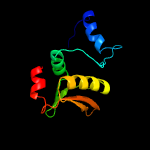
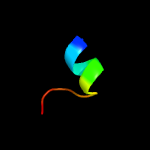
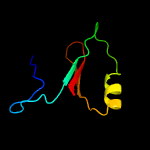
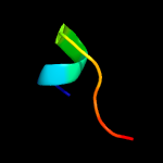
Top model
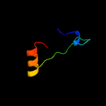
Image coloured by rainbow N → C terminus
Model dimensions (Å):X:50.884 Y:34.885 Z:35.927
Model (left) based on template c1r7mA_
Top template information
PDB header:hydrolase/dna
Chain: A: PDB Molecule:intron-encoded endonuclease i-scei;
PDBTitle: the homing endonuclease i-scei bound to its dna recognition2 region
Confidence and coverage
Confidence: 38.6% Coverage: 53%
102 residues ( 53% of your sequence) have been modelled with 38.6% confidence by the single highest scoring template.
You may wish to submit your sequence to Phyrealarm. This will automatically scan your sequence every week for new potential templates as they appear in the Phyre2 library.
Please note: You must be registered and logged in to use Phyrealarm.
3D viewing
Interactive 3D view in JSmol
For other options to view your downloaded structure offline see the FAQ

Sequence analysis 

Download FASTA version

Secondary structure and disorder prediction [Show] 

Domain analysis [Show] 

Detailed template information [Hide] 


#
Template Alignment Coverage3D Model Confidence
% i.d. Template Information
1c1r7mA_


38.6 14
PDB header:hydrolase/dna
Chain: A: PDB Molecule:intron-encoded endonuclease i-scei;
PDBTitle: the homing endonuclease i-scei bound to its dna recognition2 region
2d1dq3a4


19.6 18
Fold:Homing endonuclease-like
Superfamily:Homing endonucleases
Family:Intein endonuclease
3d1em8a_


16.7 20
Fold:DNA polymerase III chi subunit
Superfamily:DNA polymerase III chi subunit
Family:DNA polymerase III chi subunit
4d1jala2


13.9 35
Fold:beta-Grasp (ubiquitin-like)
Superfamily:TGS-like
Family:G domain-linked domain
5c4za1A_


13.4 42
PDB header:transferase
Chain: A: PDB Molecule:nosa;
PDBTitle: crystal structure of nosa involved in nosiheptide biosynthesis
6d1ni3a2


11.8 30
Fold:beta-Grasp (ubiquitin-like)
Superfamily:TGS-like
Family:G domain-linked domain
7d1jvaa3


10.2 12
Fold:Homing endonuclease-like
Superfamily:Homing endonucleases
Family:Intein endonuclease
8c2dzaA_


8.8 34
PDB header:transferase
Chain: A: PDB Molecule:dihydropteroate synthase;
PDBTitle: crystal structure of dihydropteroate synthase from thermus2 thermophilus hb8 in complex with 4-aminobenzoate
9d1k7ja_


6.3 21
Fold:YrdC/RibB
Superfamily:YrdC/RibB
Family:YrdC-like
10c3dluA_


6.2 15
PDB header:rna binding protein
Chain: A: PDB Molecule:signal recognition particle 19 kda protein;
PDBTitle: structures of srp54 and srp19, the two proteins assembling the2 ribonucleic core of the signal recognition particle from the archaeon3 pyrococcus furiosus.
11c2qsiB_


5.9 16
PDB header:structural genomics, unknown function
Chain: B: PDB Molecule:putative hydrogenase expression/formation protein hupg;
PDBTitle: crystal structure of putative hydrogenase expression/formation protein2 hupg from rhodopseudomonas palustris cga009
12d1r7ma2


5.5 15
Fold:Homing endonuclease-like
Superfamily:Homing endonucleases
Family:Group I mobile intron endonuclease
13c2faoB_


5.5 25
PDB header:hydrolase/transferase
Chain: B: PDB Molecule:probable atp-dependent dna ligase;
PDBTitle: crystal structure of pseudomonas aeruginosa ligd polymerase2 domain
14c3ccmG_


5.3 36
PDB header:ribosome
Chain: G: PDB Molecule:50s ribosomal protein l10e;
PDBTitle: structure of anisomycin resistant 50s ribosomal subunit: 23s rrna2 mutation g2611u
15c1vqkG_


5.3 36
PDB header:ribosome
Chain: G: PDB Molecule:acidic ribosomal protein p0 homolog;
PDBTitle: the structure of ccda-phe-cap-bio bound to the a site of the ribosomal2 subunit of haloarcula marismortui
16c1yitG_


5.3 36
PDB header:ribosome/antibiotic
Chain: G: PDB Molecule:acidic ribosomal protein p0 homolog;
PDBTitle: crystal structure of virginiamycin m and s bound to the 50s ribosomal2 subunit of haloarcula marismortui
17c3i56G_


5.3 36
PDB header:ribosome/antibiotic
Chain: G: PDB Molecule:50s ribosomal protein l10e;
PDBTitle: co-crystal structure of triacetyloleandomcyin bound to the large2 ribosomal subunit
18c1kc8I_


5.3 36
PDB header:ribosome
Chain: I: PDB Molecule:ribosomal protein l10;
PDBTitle: co-crystal structure of blasticidin s bound to the 50s2 ribosomal subunit
19c1k73I_


5.3 36
PDB header:ribosome
Chain: I: PDB Molecule:ribosomal protein l10;
PDBTitle: co-crystal structure of anisomycin bound to the 50s2 ribosomal subunit
20c1s72G_


5.3 36
PDB header:ribosome
Chain: G: PDB Molecule:acidic ribosomal protein p0 homolog;
PDBTitle: refined crystal structure of the haloarcula marismortui large2 ribosomal subunit at 2.4 angstrom resolution
21c1kqsG_


not modelled 5.3 36
PDB header:ribosome
Chain: G: PDB Molecule:ribosomal protein l10;
PDBTitle: the haloarcula marismortui 50s complexed with a2 pretranslocational intermediate in protein synthesis
22c1m90I_


not modelled 5.3 36
PDB header:ribosome
Chain: I: PDB Molecule:ribosomal protein l10;
PDBTitle: co-crystal structure of cca-phe-caproic acid-biotin and sparsomycin2 bound to the 50s ribosomal subunit
23c1m1kI_


not modelled 5.3 36
PDB header:ribosome
Chain: I: PDB Molecule:ribosomal protein l10;
PDBTitle: co-crystal structure of azithromycin bound to the 50s2 ribosomal subunit of haloarcula marismortui
24c1yjwG_


not modelled 5.3 36
PDB header:ribosome/antibiotic
Chain: G: PDB Molecule:acidic ribosomal protein p0 homolog;
PDBTitle: crystal structure of quinupristin bound to the g2099a mutant 50s2 ribosomal subunit of haloarcula marismortui
25c3cceG_


not modelled 5.3 36
PDB header:ribosome
Chain: G: PDB Molecule:50s ribosomal protein l10e;
PDBTitle: structure of anisomycin resistant 50s ribosomal subunit: 23s rrna2 mutation u2535a
26c1vq7G_


not modelled 5.3 36
PDB header:ribosome
Chain: G: PDB Molecule:acidic ribosomal protein p0 homolog;
PDBTitle: the structure of the transition state analogue "dca" bound to the2 large ribosomal subunit of haloarcula marismortui
27c3ccuG_


not modelled 5.3 36
PDB header:ribosome
Chain: G: PDB Molecule:50s ribosomal protein l10e;
PDBTitle: structure of anisomycin resistant 50s ribosomal subunit: 23s rrna2 mutation g2482c
28c1w2bG_


not modelled 5.3 36
PDB header:ribosome
Chain: G: PDB Molecule:50s ribosomal protein l10e;
PDBTitle: trigger factor ribosome binding domain in complex with 50s
29c1kd1I_


not modelled 5.3 36
PDB header:ribosome
Chain: I: PDB Molecule:ribosomal protein l10;
PDBTitle: co-crystal structure of spiramycin bound to the 50s2 ribosomal subunit of haloarcula marismortui
30c1qvgG_


not modelled 5.3 36
PDB header:ribosome
Chain: G: PDB Molecule:50s ribosomal protein l10e;
PDBTitle: structure of cca oligonucleotide bound to the trna binding sites of2 the large ribosomal subunit of haloarcula marismortui
31c1jj2G_


not modelled 5.3 36
PDB header:ribosome
Chain: G: PDB Molecule:ribosomal protein l10;
PDBTitle: fully refined crystal structure of the haloarcula2 marismortui large ribosomal subunit at 2.4 angstrom3 resolution
32c1k8aI_


not modelled 5.3 36
PDB header:ribosome
Chain: I: PDB Molecule:ribosomal protein l10;
PDBTitle: co-crystal structure of carbomycin a bound to the 50s2 ribosomal subunit of haloarcula marismortui
33c2otlG_


not modelled 5.3 36
PDB header:ribosome
Chain: G: PDB Molecule:50s ribosomal protein l10e;
PDBTitle: girodazole bound to the large subunit of haloarcula marismortui
34c3cc2G_


not modelled 5.3 36
PDB header:ribosome
Chain: G: PDB Molecule:50s ribosomal protein l10e;
PDBTitle: the refined crystal structure of the haloarcula marismortui large2 ribosomal subunit at 2.4 angstrom resolution with rrna sequence for3 the 23s rrna and genome-derived sequences for r-proteins
35c1k9mI_


not modelled 5.3 36
PDB header:ribosome
Chain: I: PDB Molecule:ribosomal protein l10;
PDBTitle: co-crystal structure of tylosin bound to the 50s ribosomal2 subunit of haloarcula marismortui
36c3cmaG_


not modelled 5.3 36
PDB header:ribosome
Chain: G: PDB Molecule:50s ribosomal protein l10e;
PDBTitle: the structure of cca and cca-phe-cap-bio bound to the large ribosomal2 subunit of haloarcula marismortui
37c3i55G_


not modelled 5.3 36
PDB header:ribosome/antibiotic
Chain: G: PDB Molecule:50s ribosomal protein l10e;
PDBTitle: co-crystal structure of mycalamide a bound to the large ribosomal2 subunit
38c1vqmG_


not modelled 5.3 36
PDB header:ribosome
Chain: G: PDB Molecule:acidic ribosomal protein p0 homolog;
PDBTitle: the structure of the transition state analogue "dan" bound to the2 large ribosomal subunit of haloarcula marismortui
39c1q7yI_


not modelled 5.3 36
PDB header:ribosome
Chain: I: PDB Molecule:acidic ribosomal protein p0 homolog;
PDBTitle: crystal structure of ccdap-puromycin bound at the peptidyl transferase2 center of the 50s ribosomal subunit
40c1vq5G_


not modelled 5.3 36
PDB header:ribosome
Chain: G: PDB Molecule:acidic ribosomal protein p0 homolog;
PDBTitle: the structure of the transition state analogue "raa" bound to the2 large ribosomal subunit of haloarcula marismortui
41c2otjG_


not modelled 5.3 36
PDB header:ribosome
Chain: G: PDB Molecule:50s ribosomal protein l10e;
PDBTitle: 13-deoxytedanolide bound to the large subunit of haloarcula2 marismortui
42c1qvfG_


not modelled 5.3 36
PDB header:ribosome
Chain: G: PDB Molecule:50s ribosomal protein l10e;
PDBTitle: structure of a deacylated trna minihelix bound to the e site of the2 large ribosomal subunit of haloarcula marismortui
43c1vq9G_


not modelled 5.3 36
PDB header:ribosome
Chain: G: PDB Molecule:acidic ribosomal protein p0 homolog;
PDBTitle: the structure of cca-phe-cap-bio and the antibiotic sparsomycin bound2 to the large ribosomal subunit of haloarcula marismortui
44c1yjnG_


not modelled 5.3 36
PDB header:ribosome
Chain: G: PDB Molecule:acidic ribosomal protein p0 homolog;
PDBTitle: crystal structure of clindamycin bound to the g2099a mutant 50s2 ribosomal subunit of haloarcula marismortui
45c3ow2G_


not modelled 5.3 36
PDB header:ribosome
Chain: G: PDB Molecule:50s ribosomal protein l10e;
PDBTitle: crystal structure of enhanced macrolide bound to 50s ribosomal subunit
46c1yi2G_


not modelled 5.3 36
PDB header:ribosome
Chain: G: PDB Molecule:acidic ribosomal protein p0 homolog;
PDBTitle: crystal structure of erythromycin bound to the g2099a mutant 50s2 ribosomal subunit of haloarcula marismortui
47c1vq8G_


not modelled 5.3 36
PDB header:ribosome
Chain: G: PDB Molecule:acidic ribosomal protein p0 homolog;
PDBTitle: the structure of ccda-phe-cap-bio and the antibiotic sparsomycin bound2 to the large ribosomal subunit of haloarcula marismortui
48c1q86I_


not modelled 5.3 36
PDB header:ribosome
Chain: I: PDB Molecule:acidic ribosomal protein p0 homolog;
PDBTitle: crystal structure of cca-phe-cap-biotin bound simultaneously at half2 occupancy to both the a-site and p-site of the the 50s ribosomal3 subunit.
49c3g6eG_


not modelled 5.3 36
PDB header:ribosome
Chain: G: PDB Molecule:50s ribosomal protein l10e;
PDBTitle: co-crystal structure of homoharringtonine bound to the large ribosomal2 subunit
50c1vqoG_


not modelled 5.3 36
PDB header:ribosome
Chain: G: PDB Molecule:acidic ribosomal protein p0 homolog;
PDBTitle: the structure of ccpmn bound to the large ribosomal subunit haloarcula2 marismortui
51c1vq4G_


not modelled 5.3 36
PDB header:ribosome
Chain: G: PDB Molecule:acidic ribosomal protein p0 homolog;
PDBTitle: the structure of the transition state analogue "daa" bound to the2 large ribosomal subunit of haloarcula marismortui
52c1yijG_


not modelled 5.3 36
PDB header:ribosome
Chain: G: PDB Molecule:acidic ribosomal protein p0 homolog;
PDBTitle: crystal structure of telithromycin bound to the g2099a mutant 50s2 ribosomal subunit of haloarcula marismortui
53c3cxcG_


not modelled 5.3 36
PDB header:ribosome
Chain: G: PDB Molecule:ribosomal protein l10;
PDBTitle: the structure of an enhanced oxazolidinone inhibitor bound2 to the 50s ribosomal subunit of h. marismortui
54c1q81I_


not modelled 5.3 36
PDB header:ribosome
Chain: I: PDB Molecule:acidic ribosomal protein p0 homolog;
PDBTitle: crystal structure of minihelix with 3' puromycin bound to a-site of2 the 50s ribosomal subunit.
55c1njiI_


not modelled 5.3 36
PDB header:ribosome
Chain: I: PDB Molecule:acidic ribosomal protein p0 homolog;
PDBTitle: structure of chloramphenicol bound to the 50s ribosomal2 subunit
56c3ccvG_


not modelled 5.3 36
PDB header:ribosome
Chain: G: PDB Molecule:50s ribosomal protein l10e;
PDBTitle: structure of anisomycin resistant 50s ribosomal subunit: 23s rrna2 mutation g2616a
57c3cd6G_


not modelled 5.3 36
PDB header:ribosome
Chain: G: PDB Molecule:50s ribosomal protein l10e;
PDBTitle: co-cystal of large ribosomal subunit mutant g2616a with cc-puromycin
58c1yj9G_


not modelled 5.3 36
PDB header:ribosome
Chain: G: PDB Molecule:acidic ribosomal protein p0 homolog;
PDBTitle: crystal structure of the mutant 50s ribosomal subunit of haloarcula2 marismortui containing a three residue deletion in l22
59c3cc4G_


not modelled 5.3 36
PDB header:ribosome
Chain: G: PDB Molecule:50s ribosomal protein l10e;
PDBTitle: co-crystal structure of anisomycin bound to the 50s ribosomal subunit
60c2qexG_


not modelled 5.3 36
PDB header:ribosome
Chain: G: PDB Molecule:acidic ribosomal protein p0 homolog;
PDBTitle: negamycin binds to the wall of the nascent chain exit tunnel of the2 50s ribosomal subunit
61c3cclG_


not modelled 5.3 36
PDB header:ribosome
Chain: G: PDB Molecule:50s ribosomal protein l10e;
PDBTitle: structure of anisomycin resistant 50s ribosomal subunit: 23s rrna2 mutation u2535c. density for anisomycin is visible but not included3 in model.
62c3cpwG_


not modelled 5.3 36
PDB header:ribosome
Chain: G: PDB Molecule:50s ribosomal protein l10e;
PDBTitle: the structure of the antibiotic linezolid bound to the large ribosomal2 subunit of haloarcula marismortui
63c3cc7G_


not modelled 5.3 36
PDB header:ribosome
Chain: G: PDB Molecule:50s ribosomal protein l10e;
PDBTitle: structure of anisomycin resistant 50s ribosomal subunit: 23s rrna2 mutation c2487u
64c1vq6G_


not modelled 5.3 36
PDB header:ribosome
Chain: G: PDB Molecule:acidic ribosomal protein p0 homolog;
PDBTitle: the structure of c-hpmn and cca-phe-cap-bio bound to the large2 ribosomal subunit of haloarcula marismortui
65c1q82I_


not modelled 5.3 36
PDB header:ribosome
Chain: I: PDB Molecule:acidic ribosomal protein p0 homolog;
PDBTitle: crystal structure of cc-puromycin bound to the a-site of the 50s2 ribosomal subunit
66c1n8rI_


not modelled 5.3 36
PDB header:ribosome
Chain: I: PDB Molecule:acidic ribosomal protein p0 homolog;
PDBTitle: structure of large ribosomal subunit in complex with2 virginiamycin m
67c1vqpG_


not modelled 5.3 36
PDB header:ribosome
Chain: G: PDB Molecule:acidic ribosomal protein p0 homolog;
PDBTitle: the structure of the transition state analogue "rap" bound to the2 large ribosomal subunit of haloarcula marismortui
68c1yhqG_


not modelled 5.3 36
PDB header:ribosome
Chain: G: PDB Molecule:acidic ribosomal protein p0 homolog;
PDBTitle: crystal structure of azithromycin bound to the g2099a mutant 50s2 ribosomal subunit of haloarcula marismortui
69c3ccqG_


not modelled 5.3 36
PDB header:ribosome
Chain: G: PDB Molecule:50s ribosomal protein l10e;
PDBTitle: structure of anisomycin resistant 50s ribosomal subunit: 23s rrna2 mutation a2488u
70c3ccjG_


not modelled 5.3 36
PDB header:ribosome
Chain: G: PDB Molecule:50s ribosomal protein l10e;
PDBTitle: structure of anisomycin resistant 50s ribosomal subunit: 23s rrna2 mutation c2534u
71c1vqlG_


not modelled 5.3 36
PDB header:ribosome
Chain: G: PDB Molecule:acidic ribosomal protein p0 homolog;
PDBTitle: the structure of the transition state analogue "dcsn" bound to the2 large ribosomal subunit of haloarcula marismortui
72c3g71G_


not modelled 5.3 36
PDB header:ribosome
Chain: G: PDB Molecule:50s ribosomal protein l10e;
PDBTitle: co-crystal structure of bruceantin bound to the large ribosomal2 subunit
73c3ccsG_


not modelled 5.1 33
PDB header:ribosome
Chain: G: PDB Molecule:50s ribosomal protein l10e;
PDBTitle: structure of anisomycin resistant 50s ribosomal subunit: 23s rrna2 mutation g2482a

Binding site prediction 

Due to computational demand, binding site predictions are not run for batch jobs

If you want to predict binding sites, please manually submit your model of choice to 3DLigandSite