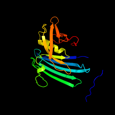
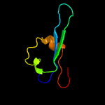
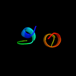
1 d2h1ta1
100.0
27
Fold: Spiral beta-rollSuperfamily: PA1994-likeFamily: PA1994-like
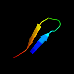
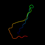
2 d1jiwi_
45.3
14
Fold: Streptavidin-likeSuperfamily: beta-Barrel protease inhibitorsFamily: Metalloprotease inhibitor
3 c5aj1A_
19.9
50
PDB header: structural proteinChain: A: PDB Molecule: swi/snf-related matrix-associated actin-dependent regulatorPDBTitle: solution structure of the smarc domain
4 c2k7mA_
14.1
28
PDB header: membrane proteinChain: A: PDB Molecule: gap junction alpha-5 protein;PDBTitle: structure of the connexin40 carboxyl terminal domain
5 c4d77A_
12.0
29
PDB header: signaling proteinChain: A: PDB Molecule: gliomedin;PDBTitle: high-resolution structure of the extracellular olfactomedin2 domain from gliomedin
6 c2gtjA_
12.0
22
PDB header: signaling proteinChain: A: PDB Molecule: fyn-binding protein;PDBTitle: reduced form of adap hsh3-n-domain
7 c6homD_
10.8
50
PDB header: gene regulationChain: D: PDB Molecule: ccr4-not transcription complex subunit 4, isoform l;PDBTitle: drosophila not4 cbm peptide bound to human caf40
8 c5tvzA_
10.7
15
PDB header: transport proteinChain: A: PDB Molecule: nucleoporin pom152;PDBTitle: solution nmr structure of saccharomyces cerevisiae pom152 ig-like2 repeat, residues 718-820
9 c5amoA_
10.6
29
PDB header: signaling proteinChain: A: PDB Molecule: noelin;PDBTitle: structure of a mouse olfactomedin-1 disulfide-linked dimer of the2 olfactomedin domain and part of the coiled coil
10 c4wxuA_
10.1
36
PDB header: protein bindingChain: A: PDB Molecule: myocilin;PDBTitle: crystal structure of the selenomthionine incorporated myocilin2 olfactomedin domain e396d variant.
11 c3m4wH_
9.9
63
PDB header: signaling protein/signaling proteinChain: H: PDB Molecule: sigma-e factor negative regulatory protein;PDBTitle: structural basis for the negative regulation of bacterial stress2 response by rseb
12 c6homB_
9.3
50
PDB header: gene regulationChain: B: PDB Molecule: ccr4-not transcription complex subunit 4, isoform l;PDBTitle: drosophila not4 cbm peptide bound to human caf40
13 c6honD_
9.3
50
PDB header: gene regulationChain: D: PDB Molecule: ccr4-not transcription complex subunit 4, isoform l;PDBTitle: drosophila not4 cbm peptide bound to human caf40
14 c2w56B_
9.2
22
PDB header: unknown functionChain: B: PDB Molecule: vc0508;PDBTitle: structure of the hypothetical protein vc0508 from vibrio cholerae2 vsp-ii pathogenicity island
15 c6honB_
9.1
50
PDB header: gene regulationChain: B: PDB Molecule: ccr4-not transcription complex subunit 4, isoform l;PDBTitle: drosophila not4 cbm peptide bound to human caf40
16 c6eluJ_
8.9
27
PDB header: antitoxinChain: J: PDB Molecule: serum resistance associated; vsg protein;PDBTitle: structure of serum resistance associated protein from t. b.2 rhodesiense
17 d1jz8a4
8.8
18
Fold: SupersandwichSuperfamily: Galactose mutarotase-likeFamily: beta-Galactosidase, domain 5
18 d2d81a1
8.7
36
Fold: alpha/beta-HydrolasesSuperfamily: alpha/beta-HydrolasesFamily: PHB depolymerase-like
19 d1o1za_
8.0
36
Fold: TIM beta/alpha-barrelSuperfamily: PLC-like phosphodiesterasesFamily: Glycerophosphoryl diester phosphodiesterase
20 d3bb9a1
7.9
9
Fold: Cystatin-likeSuperfamily: NTF2-likeFamily: SO0125-like
21 c6nk6G_
not modelled
7.9
16
PDB header: virus like particle/signaling proteinChain: G: PDB Molecule: e2 glycoprotein;PDBTitle: electron cryo-microscopy of chikungunya vlp in complex with mouse2 mxra8 receptor
22 c1wx4B_
not modelled
7.9
25
PDB header: oxidoreductase/metal transportChain: B: PDB Molecule: melc;PDBTitle: crystal structure of the oxy-form of the copper-bound streptomyces2 castaneoglobisporus tyrosinase complexed with a caddie protein3 prepared by the addition of dithiothreitol
23 c5afbA_
not modelled
7.8
36
PDB header: signaling proteinChain: A: PDB Molecule: latrophilin-3;PDBTitle: crystal structure of the latrophilin3 lectin and2 olfactomedin domains
24 c2mpvA_
not modelled
7.3
27
PDB header: protein bindingChain: A: PDB Molecule: major fimbrial subunit of aggregative adherence fimbria iiPDBTitle: structural insight into host recognition and biofilm formation by2 aggregative adherence fimbriae of enteroaggregative esherichia coli
25 d1l5aa2
not modelled
7.2
33
Fold: CoA-dependent acyltransferasesSuperfamily: CoA-dependent acyltransferasesFamily: NRPS condensation domain (amide synthase)
26 c3rlgA_
not modelled
7.0
29
PDB header: hydrolaseChain: A: PDB Molecule: sphingomyelin phosphodiesterase d lisictox-alphaia1a;PDBTitle: crystal structure of loxosceles intermedia phospholipase d isoform 12 h12a mutant
27 d2q07a3
not modelled
6.6
57
Fold: Cystatin-likeSuperfamily: Pre-PUA domainFamily: AF0587 pre C-terminal domain-like
28 c3j0cH_
not modelled
6.5
15
PDB header: virusChain: H: PDB Molecule: e2 envelope glycoprotein;PDBTitle: models of e1, e2 and cp of venezuelan equine encephalitis virus tc-832 strain restrained by a near atomic resolution cryo-em map
29 c3n43B_
not modelled
6.3
15
PDB header: viral proteinChain: B: PDB Molecule: e2 envelope glycoprotein;PDBTitle: crystal structures of the mature envelope glycoprotein complex2 (trypsin cleavage) of chikungunya virus.
30 c5u9oD_
not modelled
6.3
14
PDB header: cell cycleChain: D: PDB Molecule: plastid division protein cdp1, chloroplastic,plastidPDBTitle: cocrystal structure of the intermembrane space region of the plastid2 division proteins parc6 and pdv1
31 c2xfbI_
not modelled
6.2
15
PDB header: virusChain: I: PDB Molecule: e2 envelope glycoprotein;PDBTitle: the chikungunya e1 e2 envelope glycoprotein complex fit into2 the sindbis virus cryo-em map
32 c6g1cV_
not modelled
5.9
20
PDB header: antitoxinChain: V: PDB Molecule: antitoxin hicb;PDBTitle: crystal structure of the n-terminal domain of burkholderia2 pseudomallei antitoxin hicb
33 c2r9qD_
not modelled
5.9
35
PDB header: hydrolaseChain: D: PDB Molecule: 2'-deoxycytidine 5'-triphosphate deaminase;PDBTitle: crystal structure of 2'-deoxycytidine 5'-triphosphate deaminase from2 agrobacterium tumefaciens
34 c4q6xA_
not modelled
5.8
29
PDB header: lyaseChain: A: PDB Molecule: phospholipase d stsictox-betaic1;PDBTitle: structure of phospholipase d beta1b1i from sicarius terrosus venom at2 2.14 a resolution
35 c2f9rC_
not modelled
5.7
29
PDB header: hydrolaseChain: C: PDB Molecule: sphingomyelinase d 1;PDBTitle: crystal structure of the inactive state of the smase i, a2 sphingomyelinase d from loxosceles laeta venom
36 d1yb3a1
not modelled
5.4
12
Fold: YktB/PF0168-likeSuperfamily: YktB/PF0168-likeFamily: PF0168-like
37 c4hvmC_
not modelled
5.4
11
PDB header: biosynthetic proteinChain: C: PDB Molecule: tlmii;PDBTitle: crystal structure of tallysomycin biosynthesis protein tlmii
38 c3fzeA_
not modelled
5.3
36
PDB header: protein bindingChain: A: PDB Molecule: protein ste5;PDBTitle: structure of the 'minimal scaffold' (ms) domain of ste5 that2 cocatalyzes fus3 phosphorylation by ste7
39 d1vd6a1
not modelled
5.2
36
Fold: TIM beta/alpha-barrelSuperfamily: PLC-like phosphodiesterasesFamily: Glycerophosphoryl diester phosphodiesterase