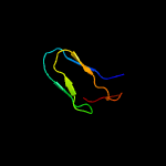
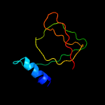
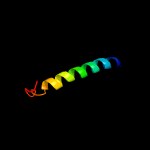
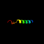
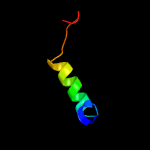
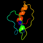
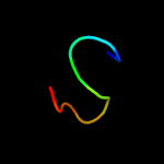
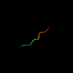
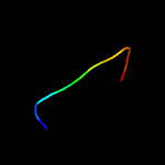
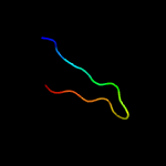
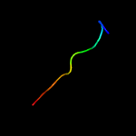
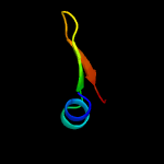
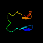
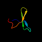
1 c5klcA_
47.8
33
PDB header: sugar binding proteinChain: A: PDB Molecule: carbohydrate binding module e1;PDBTitle: structure of cbm_e1, a novel carbohydrate-binding module found by2 sugar cane soil metagenome
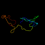
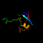
2 c4nl6C_
28.9
13
PDB header: splicingChain: C: PDB Molecule: survival motor neuron protein;PDBTitle: structure of the full-length form of the protein smn found in healthy2 patients
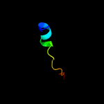
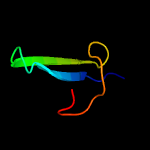
3 c4a1sE_
26.9
44
PDB header: cell cycleChain: E: PDB Molecule: re60102p;PDBTitle: crystallographic structure of the pins:insc complex
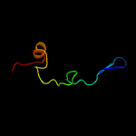
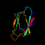
4 c3lybC_
23.6
23
PDB header: hydrolaseChain: C: PDB Molecule: putative endoribonuclease;PDBTitle: structure of putative endoribonuclease(kp1_3112) from klebsiella2 pneumoniae
5 c3gjbA_
17.9
9
PDB header: biosynthetic proteinChain: A: PDB Molecule: cytc3;PDBTitle: cytc3 with fe(ii) and alpha-ketoglutarate
6 c4pmkA_
17.8
30
PDB header: plant proteinChain: A: PDB Molecule: kiwellin;PDBTitle: crystal structure of kiwellin
7 c2lcdA_
15.6
19
PDB header: transcriptionChain: A: PDB Molecule: at-rich interactive domain-containing protein 4a;PDBTitle: solution structure of rbbp1 tudor domain
8 c5jqyA_
13.7
18
PDB header: oxidoreductaseChain: A: PDB Molecule: aspartyl/asparaginyl beta-hydroxylase;PDBTitle: aspartyl/asparaginyl beta-hydroxylase (asph)oxygenase and tpr domains2 in complex with manganese, n-oxalylglycine and factor x substrate3 peptide fragment(39mer-4ser)
9 c2momC_
13.6
30
PDB header: membrane proteinChain: C: PDB Molecule: lysosome-associated membrane glycoprotein 2;PDBTitle: structural insights of tm domain of lamp-2a in dpc micelles
10 c2momB_
12.4
30
PDB header: membrane proteinChain: B: PDB Molecule: lysosome-associated membrane glycoprotein 2;PDBTitle: structural insights of tm domain of lamp-2a in dpc micelles
11 c4d0kC_
10.3
15
PDB header: gene regulationChain: C: PDB Molecule: pab-dependent poly(a)-specific ribonuclease subunitPDBTitle: complex of chaetomium thermophilum pan2 (wd40-cs1) with pan3 (c-term)
12 c4a1aM_
10.3
11
PDB header: ribosomeChain: M: PDB Molecule: 60s ribosomal protein l5;PDBTitle: t.thermophila 60s ribosomal subunit in complex with2 initiation factor 6. this file contains 5s rrna,3 5.8s rrna and proteins of molecule 3.
13 d2fsqa1
9.9
36
Fold: LigT-likeSuperfamily: LigT-likeFamily: Atu0111-like
14 d1zxqa2
9.6
50
Fold: Immunoglobulin-like beta-sandwichSuperfamily: ImmunoglobulinFamily: I set domains
15 d1s6la2
9.4
20
Fold: NosL/MerB-likeSuperfamily: NosL/MerB-likeFamily: MerB-like
16 c5iwzA_
9.3
27
PDB header: cell cycleChain: A: PDB Molecule: synaptonemal complex protein 2;PDBTitle: synaptonemal complex protein
17 c5ue8B_
9.0
55
PDB header: exocytosisChain: B: PDB Molecule: protein unc-13 homolog a;PDBTitle: the crystal structure of munc13-1 c1c2bmun domain
18 c4dzoA_
8.8
17
PDB header: cell cycleChain: A: PDB Molecule: mitotic spindle assembly checkpoint protein mad1;PDBTitle: structure of human mad1 c-terminal domain reveals its involvement in2 kinetochore targeting
19 d1wyka_
8.4
19
Fold: Trypsin-like serine proteasesSuperfamily: Trypsin-like serine proteasesFamily: Viral proteases
20 d1e8qa_
8.2
21
Fold: Cellulose docking domain, dockeringSuperfamily: Cellulose docking domain, dockeringFamily: Cellulose docking domain, dockering
21 d1ep5a_
not modelled
8.2
22
Fold: Trypsin-like serine proteasesSuperfamily: Trypsin-like serine proteasesFamily: Viral proteases
22 c3hqxA_
not modelled
8.0
22
PDB header: structural genomics, unknown functionChain: A: PDB Molecule: upf0345 protein aciad0356;PDBTitle: crystal structure of protein of unknown function (duf1255,pf06865)2 from acinetobacter sp. adp1
23 c1kxfA_
not modelled
7.9
19
PDB header: viral proteinChain: A: PDB Molecule: sindbis virus capsid protein;PDBTitle: sindbis virus capsid, (wild-type) residues 1-264, tetragonal crystal2 form (form ii)
24 c2yewG_
not modelled
7.7
25
PDB header: virusChain: G: PDB Molecule: capsid protein;PDBTitle: modeling barmah forest virus structural proteins
25 c1t0pB_
not modelled
7.7
50
PDB header: immune systemChain: B: PDB Molecule: intercellular adhesion molecule-3;PDBTitle: structural basis of icam recognition by integrin alpahlbeta2 revealed2 in the complex structure of binding domains of icam-3 and alphalbeta23 at 1.65 a
26 d1vcpa_
not modelled
7.7
25
Fold: Trypsin-like serine proteasesSuperfamily: Trypsin-like serine proteasesFamily: Viral proteases
27 c4f5cF_
not modelled
7.5
20
PDB header: hydrolase/viral proteinChain: F: PDB Molecule: prcv spike protein;PDBTitle: crystal structure of the spike receptor binding domain of a porcine2 respiratory coronavirus in complex with the pig aminopeptidase n3 ectodomain
28 c2n4pA_
not modelled
7.4
19
PDB header: dna binding proteinChain: A: PDB Molecule: tar dna-binding protein 43;PDBTitle: solution structure of the n-terminal domain of tdp-43
29 c2q49B_
not modelled
6.7
21
PDB header: oxidoreductaseChain: B: PDB Molecule: probable n-acetyl-gamma-glutamyl-phosphate reductase;PDBTitle: ensemble refinement of the protein crystal structure of gene product2 from arabidopsis thaliana at2g19940
30 c5mrgA_
not modelled
6.6
19
PDB header: dna binding proteinChain: A: PDB Molecule: tar dna-binding protein 43;PDBTitle: solution structure of tdp-43 (residues 1-102)
31 c3d2hA_
not modelled
6.5
24
PDB header: oxidoreductaseChain: A: PDB Molecule: berberine bridge-forming enzyme;PDBTitle: structure of berberine bridge enzyme from eschscholzia californica,2 monoclinic crystal form
32 c2m6oA_
not modelled
6.0
43
PDB header: transcriptionChain: A: PDB Molecule: uncharacterized protein;PDBTitle: the actinobacterial transcription factor rbpa binds to the principal2 sigma subunit of rna polymerase
33 c5mdxx_
not modelled
5.9
30
PDB header: photosynthesisChain: X: PDB Molecule: photosystem ii reaction center protein x;PDBTitle: cryo-em structure of the psii supercomplex from arabidopsis thaliana
34 c5mdxX_
not modelled
5.9
30
PDB header: photosynthesisChain: X: PDB Molecule: photosystem ii reaction center protein x;PDBTitle: cryo-em structure of the psii supercomplex from arabidopsis thaliana
35 c6mx7C_
not modelled
5.9
22
PDB header: virusChain: C: PDB Molecule: capsid;PDBTitle: cryoem structure of chimeric eastern equine encephalitis virus:2 genome-binding capsid n-terminal domain
36 c3j3bD_
not modelled
5.9
17
PDB header: ribosomeChain: D: PDB Molecule: 60s ribosomal protein l5;PDBTitle: structure of the human 60s ribosomal proteins
37 c2miiA_
not modelled
5.4
21
PDB header: protein bindingChain: A: PDB Molecule: penicillin-binding protein activator lpob;PDBTitle: nmr structure of e. coli lpob
38 c4c8hC_
not modelled
5.1
23
PDB header: dna replicationChain: C: PDB Molecule: ctf4;PDBTitle: crystal structure of the c-terminal region of yeast ctf4,2 selenomethionine protein.
39 c6d6vF_
not modelled
5.0
38
PDB header: replicationChain: F: PDB Molecule: telomerase holoenzyme teb heterotrimer teb3 subunit;PDBTitle: cryoem structure of tetrahymena telomerase with telomeric dna at 4.82 angstrom resolution
40 c3jcux_
not modelled
5.0
35
PDB header: membrane proteinChain: X: PDB Molecule: photosystem ii reaction center x protein;PDBTitle: cryo-em structure of spinach psii-lhcii supercomplex at 3.2 angstrom2 resolution


