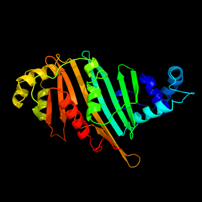
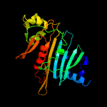
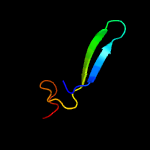
PDB header:protein transport Chain: C: PDB Molecule:espg5; PDBTitle: structure of the mycobacterium tuberculosis type vii secretion system2 chaperone espg5 in complex with pe25-ppe41 dimer
Confidence and coverage
Confidence:
100.0%
Coverage:
90%
249 residues ( 90% of your sequence) have been modelled with 100.0% confidence by the single highest scoring template.
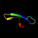
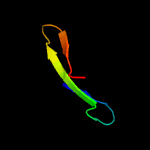
Region: 2 - 270 Aligned: 256 Modelled: 269 Confidence: 100.0% Identity: 25% PDB header:protein transport Chain: B: PDB Molecule:espg3; PDBTitle: structure of espg3 chaperone from the type vii (esx-3) secretion2 system
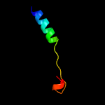
Region: 2 - 270 Aligned: 237 Modelled: 269 Confidence: 100.0% Identity: 27% PDB header:chaperone, hydrolase Chain: A: PDB Molecule:lysozyme, esx-1 secretion-associated protein espg1 chimera; PDBTitle: structure of espg1 chaperone from the type vii (esx-1) secretion2 system determined with the assistance of n-terminal t4 lysozyme3 fusion
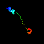
Region: 2 - 265 Aligned: 252 Modelled: 264 Confidence: 100.0% Identity: 23% PDB header:protein transport Chain: A: PDB Molecule:esx-3 secretion-associated protein espg3; PDBTitle: crystal structure of espg3 from the esx-3 type vii secretion system of2 m. tuberculosis
Region: 2 - 270 Aligned: 227 Modelled: 239 Confidence: 100.0% Identity: 26% PDB header:chaperone Chain: B: PDB Molecule:espg3; PDBTitle: structure of espg3 chaperone from the type vii (esx-3) secretion2 system, space group p43212
Region: 1 - 21 Aligned: 21 Modelled: 21 Confidence: 7.2% Identity: 24% PDB header:cell adhesion Chain: B: PDB Molecule:amyloid beta a4 precursor protein-binding family b member PDBTitle: vinculin head (1-258) in complex with a riam fragment