| 1 |
|
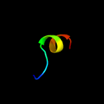
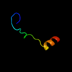
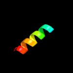
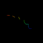
PDB 2vs0 chain B
Region: 17 - 27
Aligned: 11
Modelled: 11
Confidence: 24.2%
Identity: 36%
PDB header:cell invasion
Chain: B: PDB Molecule:virulence factor esxa;
PDBTitle: structural analysis of homodimeric staphylococcal aureus2 virulence factor esxa
Phyre2
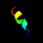
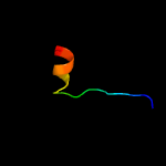
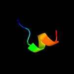
| 2 |
|
PDB 6joo chain A
Region: 77 - 87
Aligned: 11
Modelled: 11
Confidence: 16.0%
Identity: 73%
PDB header:hydrolase/dna/rna
Chain: A: PDB Molecule:crispr-associated protein,crispr-associated endonuclease
PDBTitle: crystal structure of corynebacterium diphtheriae cas9 in complex with2 sgrna and target dna
Phyre2
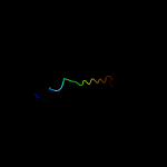
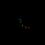
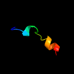
| 3 |
|
PDB 6gyg chain A
Region: 17 - 27
Aligned: 11
Modelled: 11
Confidence: 15.3%
Identity: 36%
PDB header:transcription
Chain: A: PDB Molecule:transcription regulator reg576;
PDBTitle: x-ray structure of the apo form of the establishement gene regulator2 reg576 of the g+ plasmid p576
Phyre2
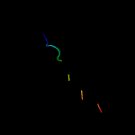
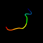
| 4 |
|
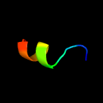
PDB 6a0c chain B
Region: 30 - 49
Aligned: 20
Modelled: 20
Confidence: 12.4%
Identity: 35%
PDB header:structural protein
Chain: B: PDB Molecule:collagen type iii peptide;
PDBTitle: structure of a triple-helix region of human collagen type iii
Phyre2
| 5 |
|
PDB 5zbi chain B
Region: 5 - 30
Aligned: 26
Modelled: 26
Confidence: 12.2%
Identity: 38%
PDB header:plant protein
Chain: B: PDB Molecule:peptide asparaginyl ligase;
PDBTitle: crystal structure of asparaginyl endopeptidases from viola canadensis
Phyre2
| 6 |
|
PDB 6a0c chain C
Region: 30 - 49
Aligned: 20
Modelled: 20
Confidence: 11.6%
Identity: 35%
PDB header:structural protein
Chain: C: PDB Molecule:collagen type iii peptide;
PDBTitle: structure of a triple-helix region of human collagen type iii
Phyre2
| 7 |
|
PDB 6a0c chain A
Region: 30 - 49
Aligned: 20
Modelled: 20
Confidence: 11.6%
Identity: 35%
PDB header:structural protein
Chain: A: PDB Molecule:collagen type iii peptide;
PDBTitle: structure of a triple-helix region of human collagen type iii
Phyre2
| 8 |
|
PDB 4iog chain D
Region: 17 - 27
Aligned: 11
Modelled: 11
Confidence: 11.3%
Identity: 55%
PDB header:unknown function
Chain: D: PDB Molecule:secreted protein esxb;
PDBTitle: the crystal structure of a secreted protein esxb (wild-type, in p212 space group) from bacillus anthracis str. sterne
Phyre2
| 9 |
|
PDB 3ter chain A
Region: 69 - 86
Aligned: 18
Modelled: 18
Confidence: 10.1%
Identity: 17%
PDB header:metal binding protein
Chain: A: PDB Molecule:mammalian stromal interaction molecule-1;
PDBTitle: crystal structure of soar domain with inhibition helix from c. elegans
Phyre2
| 10 |
|
PDB 4fgu chain B
Region: 13 - 30
Aligned: 18
Modelled: 18
Confidence: 9.2%
Identity: 50%
PDB header:hydrolase
Chain: B: PDB Molecule:legumain;
PDBTitle: crystal structure of prolegumain
Phyre2
| 11 |
|
PDB 6hg7 chain C
Region: 1 - 14
Aligned: 14
Modelled: 14
Confidence: 9.1%
Identity: 64%
PDB header:structural protein
Chain: C: PDB Molecule:collagen alpha-1(ii) chain;
PDBTitle: crystal structure of a collagen ii fragment containing the binding2 site of pedf and comp, (pog)4-lkg hrg ftg lqg-pog(4)
Phyre2
| 12 |
|
PDB 6hg7 chain A
Region: 1 - 14
Aligned: 14
Modelled: 14
Confidence: 9.1%
Identity: 64%
PDB header:structural protein
Chain: A: PDB Molecule:collagen alpha-1(ii) chain;
PDBTitle: crystal structure of a collagen ii fragment containing the binding2 site of pedf and comp, (pog)4-lkg hrg ftg lqg-pog(4)
Phyre2
| 13 |
|
PDB 6hg7 chain B
Region: 1 - 14
Aligned: 14
Modelled: 14
Confidence: 9.1%
Identity: 64%
PDB header:structural protein
Chain: B: PDB Molecule:collagen alpha-1(ii) chain;
PDBTitle: crystal structure of a collagen ii fragment containing the binding2 site of pedf and comp, (pog)4-lkg hrg ftg lqg-pog(4)
Phyre2
| 14 |
|
PDB 3gvm chain A
Region: 17 - 27
Aligned: 11
Modelled: 11
Confidence: 8.8%
Identity: 55%
PDB header:viral protein
Chain: A: PDB Molecule:putative uncharacterized protein sag1039;
PDBTitle: structure of the homodimeric wxg-100 family protein from streptococcus2 agalactiae
Phyre2
| 15 |
|
PDB 3teq chain B
Region: 69 - 86
Aligned: 18
Modelled: 18
Confidence: 6.7%
Identity: 33%
PDB header:signaling protein
Chain: B: PDB Molecule:stromal interaction molecule 1;
PDBTitle: crystal structure of soar domain
Phyre2
| 16 |
|
PDB 1rqb chain A domain 1
Region: 14 - 21
Aligned: 8
Modelled: 8
Confidence: 5.7%
Identity: 63%
Fold: RuvA C-terminal domain-like
Superfamily: post-HMGL domain-like
Family: Conserved carboxylase domain
Phyre2
| 17 |
|
PDB 5wsx chain A
Region: 1 - 29
Aligned: 18
Modelled: 29
Confidence: 5.3%
Identity: 61%
PDB header:hydrolase
Chain: A: PDB Molecule:uncharacterized protein;
PDBTitle: the crystal structure of sav606
Phyre2