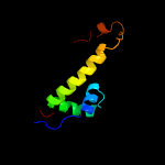
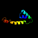
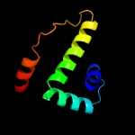
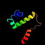
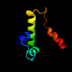
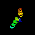
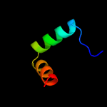
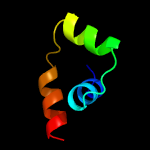
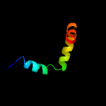
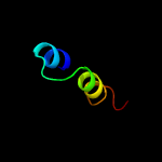
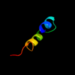
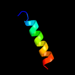
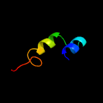
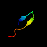
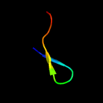
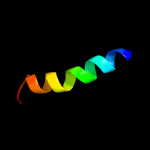
1 c2z2sD_
99.3
16
PDB header: transcriptionChain: D: PDB Molecule: anti-sigma factor chrr, transcriptional activator chrr;PDBTitle: crystal structure of rhodobacter sphaeroides sige in complex with the2 anti-sigma chrr
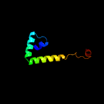
2 c5wuqD_
99.2
6
PDB header: metal binding proteinChain: D: PDB Molecule: anti-sigma-w factor rsiw;PDBTitle: crystal structure of sigw in complex with its anti-sigma rsiw, a zinc2 binding form
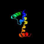
3 c3hugJ_
99.1
13
PDB header: transcription/membrane proteinChain: J: PDB Molecule: probable conserved membrane protein;PDBTitle: crystal structure of mycobacterium tuberculosis anti-sigma factor rsla2 in complex with -35 promoter binding domain of sigl
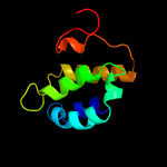
4 c5frhA_
99.1
10
PDB header: transcriptionChain: A: PDB Molecule: anti-sigma factor rsra;PDBTitle: solution structure of oxidised rsra
5 c3vdoB_
98.9
29
PDB header: dna binding protein/protein bindingChain: B: PDB Molecule: anti-sigma-k factor rska;PDBTitle: structure of extra-cytoplasmic function(ecf) sigma factor sigk in2 complex with its negative regulator rska from mycobacterium3 tuberculosis
6 c6in7A_
98.1
12
PDB header: transcriptionChain: A: PDB Molecule: sigma factor algu negative regulatory protein;PDBTitle: crystal structure of algu in complex with muca(cyto)
7 c1or7C_
95.9
16
PDB header: transcriptionChain: C: PDB Molecule: sigma-e factor negative regulatory protein;PDBTitle: crystal structure of escherichia coli sigmae with the cytoplasmic2 domain of its anti-sigma rsea
8 d1or7c_
95.9
16
Fold: N-terminal, cytoplasmic domain of anti-sigmaE factor RseASuperfamily: N-terminal, cytoplasmic domain of anti-sigmaE factor RseAFamily: N-terminal, cytoplasmic domain of anti-sigmaE factor RseA
9 c5camC_
95.8
15
PDB header: transcriptionChain: C: PDB Molecule: pupr protein;PDBTitle: crystal structure of the cytoplasmic domain of the pseudomonas putida2 anti-sigma factor pupr (semet)
10 c4ynhA_
74.9
26
PDB header: structural proteinChain: A: PDB Molecule: spindle assembly abnormal protein 5;PDBTitle: structure of the c. elegans sas-5 implico dimerization domain
11 c5m45I_
28.5
20
PDB header: ligaseChain: I: PDB Molecule: acetone carboxylase gamma subunit;PDBTitle: structure of acetone carboxylase purified from xanthobacter2 autotrophicus
12 c3kb4D_
18.5
20
PDB header: structural genomics, unknown functionChain: D: PDB Molecule: alr8543 protein;PDBTitle: crystal structure of the alr8543 protein in complex with2 geranylgeranyl monophosphate and magnesium ion from nostoc sp. pcc3 7120, northeast structural genomics consortium target nsr141
13 c5oynB_
18.3
22
PDB header: lyaseChain: B: PDB Molecule: dehydratase, ilvd/edd family;PDBTitle: crystal structure of d-xylonate dehydratase in holo-form
14 c5ze4A_
16.7
19
PDB header: lyaseChain: A: PDB Molecule: dihydroxy-acid dehydratase, chloroplastic;PDBTitle: the structure of holo- structure of dhad complex with [2fe-2s] cluster
15 c6ovtD_
12.0
22
PDB header: lyaseChain: D: PDB Molecule: dihydroxy-acid dehydratase;PDBTitle: crystal structure of ilvd from mycobacterium tuberculosis
16 d2f8aa1
11.6
10
Fold: Thioredoxin foldSuperfamily: Thioredoxin-likeFamily: Glutathione peroxidase-like
17 c5j84A_
11.4
11
PDB header: lyaseChain: A: PDB Molecule: dihydroxy-acid dehydratase;PDBTitle: crystal structure of l-arabinonate dehydratase in holo-form
18 d1igqa_
11.0
38
Fold: SH3-like barrelSuperfamily: C-terminal domain of transcriptional repressorsFamily: Transcriptional repressor protein KorB
19 d1igub_
9.9
38
Fold: SH3-like barrelSuperfamily: C-terminal domain of transcriptional repressorsFamily: Transcriptional repressor protein KorB
20 c2qsiB_
9.8
15
PDB header: structural genomics, unknown functionChain: B: PDB Molecule: putative hydrogenase expression/formation protein hupg;PDBTitle: crystal structure of putative hydrogenase expression/formation protein2 hupg from rhodopseudomonas palustris cga009
21 d1v58a1
not modelled
9.7
13
Fold: Thioredoxin foldSuperfamily: Thioredoxin-likeFamily: DsbC/DsbG C-terminal domain-like
22 c5lbmD_
not modelled
9.3
17
PDB header: transcriptionChain: D: PDB Molecule: transcriptional repressor frmr;PDBTitle: the asymmetric tetrameric structure of the formaldehyde sensing2 transcriptional repressor frmr from escherichia coli
23 c3gv1A_
not modelled
9.2
7
PDB header: structural genomics, unknown functionChain: A: PDB Molecule: disulfide interchange protein;PDBTitle: crystal structure of disulfide interchange protein from neisseria2 gonorrhoeae
24 d2cwla1
not modelled
9.1
21
Fold: Ferritin-likeSuperfamily: Ferritin-likeFamily: Manganese catalase (T-catalase)
25 d2nrka1
not modelled
8.4
19
Fold: NucleotidyltransferaseSuperfamily: NucleotidyltransferaseFamily: GrpB-like
26 d1j08a1
not modelled
8.2
21
Fold: Thioredoxin foldSuperfamily: Thioredoxin-likeFamily: PDI-like
27 d1z6ma1
not modelled
8.1
9
Fold: Thioredoxin foldSuperfamily: Thioredoxin-likeFamily: DsbA-like
28 c2mjlA_
not modelled
7.3
15
PDB header: hydrolaseChain: A: PDB Molecule: peptidyl-trna hydrolase;PDBTitle: solution structure of peptidyl-trna hyrolase from vibrio cholerae
29 d1z6na1
not modelled
7.0
30
Fold: Thioredoxin foldSuperfamily: Thioredoxin-likeFamily: Thioltransferase
30 d1t3ba1
not modelled
6.7
6
Fold: Thioredoxin foldSuperfamily: Thioredoxin-likeFamily: DsbC/DsbG C-terminal domain-like
31 c4rs7R_
not modelled
6.7
21
PDB header: dna binding proteinChain: R: PDB Molecule: parb-c;PDBTitle: structure of pnob8 parb-c
32 c2qv8B_
not modelled
6.5
4
PDB header: transport proteinChain: B: PDB Molecule: general secretion pathway protein h;PDBTitle: structure of the minor pseudopilin epsh from the type 2 secretion2 system of vibrio cholerae
33 c2odkD_
not modelled
6.5
17
PDB header: structural genomics, unknown functionChain: D: PDB Molecule: hypothetical protein;PDBTitle: putative prevent-host-death protein from nitrosomonas europaea
34 d1ftra1
not modelled
6.4
39
Fold: Ferredoxin-likeSuperfamily: Formylmethanofuran:tetrahydromethanopterin formyltransferaseFamily: Formylmethanofuran:tetrahydromethanopterin formyltransferase
35 c5lcyD_
not modelled
6.0
16
PDB header: transcriptionChain: D: PDB Molecule: frmr;PDBTitle: formaldehyde-responsive regulator frmr e64h variant from salmonella2 enterica serovar typhimurium
36 c1wx4B_
not modelled
5.8
8
PDB header: oxidoreductase/metal transportChain: B: PDB Molecule: melc;PDBTitle: crystal structure of the oxy-form of the copper-bound streptomyces2 castaneoglobisporus tyrosinase complexed with a caddie protein3 prepared by the addition of dithiothreitol
37 c2hh7A_
not modelled
5.5
13
PDB header: unknown functionChain: A: PDB Molecule: hypothetical protein csor;PDBTitle: crystal structure of cu(i) bound csor from mycobacterium tuberculosis.
38 c3tsaA_
not modelled
5.2
18
PDB header: transferaseChain: A: PDB Molecule: ndp-rhamnosyltransferase;PDBTitle: spinosyn rhamnosyltransferase spng


