| 1 |
|
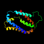
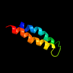
PDB 5vkv chain A
Region: 21 - 195
Aligned: 172
Modelled: 175
Confidence: 99.0%
Identity: 14%
PDB header:oxidoreductase
Chain: A: PDB Molecule:cytochrome c-type biogenesis protein ccda;
PDBTitle: solution nmr structure of the membrane electron transporter ccda
Phyre2
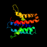
| 2 |
|
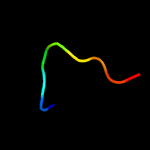
PDB 2n4x chain A
Region: 21 - 189
Aligned: 158
Modelled: 169
Confidence: 96.7%
Identity: 10%
PDB header:membrane protein
Chain: A: PDB Molecule:cytochrome c-type biogenesis protein (ccda);
PDBTitle: structure of the transmembrane electron transporter ccda
Phyre2
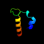
| 3 |
|
PDB 5xyv chain C
Region: 74 - 112
Aligned: 33
Modelled: 39
Confidence: 41.1%
Identity: 9%
PDB header:protein binding
Chain: C: PDB Molecule:protein deadlock;
PDBTitle: crystal structure of drosophila melanogaster rhino chromoshadow domain2 in complex with deadlock n-terminal domain
Phyre2
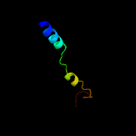
| 4 |
|
PDB 2knc chain A
Region: 55 - 92
Aligned: 38
Modelled: 38
Confidence: 12.1%
Identity: 16%
PDB header:cell adhesion
Chain: A: PDB Molecule:integrin alpha-iib;
PDBTitle: platelet integrin alfaiib-beta3 transmembrane-cytoplasmic2 heterocomplex
Phyre2
| 5 |
|
PDB 2lkg chain A
Region: 104 - 151
Aligned: 48
Modelled: 48
Confidence: 7.8%
Identity: 8%
PDB header:signaling protein
Chain: A: PDB Molecule:acetylcholine receptor;
PDBTitle: wsa major conformation
Phyre2
| 6 |
|
PDB 1px5 chain A
Region: 190 - 198
Aligned: 9
Modelled: 9
Confidence: 5.6%
Identity: 56%
PDB header:transferase
Chain: A: PDB Molecule:2'-5'-oligoadenylate synthetase 1;
PDBTitle: crystal structure of the 2'-specific and double-stranded2 rna-activated interferon-induced antiviral protein 2'-5'-3 oligoadenylate synthetase
Phyre2
| 7 |
|
PDB 1px5 chain A domain 1
Region: 190 - 198
Aligned: 9
Modelled: 9
Confidence: 5.3%
Identity: 56%
Fold: PAP/OAS1 substrate-binding domain
Superfamily: PAP/OAS1 substrate-binding domain
Family: 2'-5'-oligoadenylate synthetase 1, OAS1, second domain
Phyre2