| 1 |
|
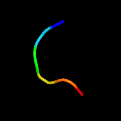
PDB 5wkn chain D
Region: 138 - 143
Aligned: 6
Modelled: 6
Confidence: 17.2%
Identity: 100%
PDB header:viral protein
Chain: D: PDB Molecule:phosphoprotein;
PDBTitle: crystal structure of the parainfluenza virus 5 nucleoprotein-2 phosphoprotein complex
Phyre2
| 2 |
|
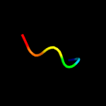
PDB 5wkn chain C
Region: 138 - 143
Aligned: 6
Modelled: 6
Confidence: 16.6%
Identity: 100%
PDB header:viral protein
Chain: C: PDB Molecule:phosphoprotein;
PDBTitle: crystal structure of the parainfluenza virus 5 nucleoprotein-2 phosphoprotein complex
Phyre2
| 3 |
|
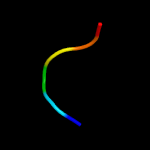
PDB 1qcr chain I
Region: 52 - 72
Aligned: 21
Modelled: 21
Confidence: 10.7%
Identity: 48%
PDB header:oxidoreductase
Chain: I: PDB Molecule:ubiquinol cytochrome c oxidoreductase;
PDBTitle: crystal structure of bovine mitochondrial cytochrome bc12 complex, alpha carbon atoms only
Phyre2
| 4 |
|
PDB 4wlp chain B
Region: 134 - 153
Aligned: 20
Modelled: 20
Confidence: 9.0%
Identity: 40%
PDB header:protein binding
Chain: B: PDB Molecule:nuclear factor related to kappa-b-binding protein;
PDBTitle: crystal structure of uch37-nfrkb inhibited deubiquitylating complex
Phyre2
| 5 |
|
PDB 5ejo chain A
Region: 97 - 114
Aligned: 18
Modelled: 18
Confidence: 7.4%
Identity: 33%
PDB header:nuclear protein
Chain: A: PDB Molecule:chromatin assembly factor 1 subunit p90;
PDBTitle: crystal structure of the winged helix domain in chromatin assembly2 factor 1 subunit p90
Phyre2
| 6 |
|
PDB 3dkb chain A
Region: 125 - 140
Aligned: 16
Modelled: 16
Confidence: 7.3%
Identity: 38%
PDB header:hydrolase
Chain: A: PDB Molecule:tumor necrosis factor, alpha-induced protein 3;
PDBTitle: crystal structure of a20, 2.5 angstrom
Phyre2
| 7 |
|
PDB 5lrv chain A
Region: 125 - 140
Aligned: 16
Modelled: 16
Confidence: 7.2%
Identity: 44%
PDB header:hydrolase
Chain: A: PDB Molecule:otu domain-containing protein 7b;
PDBTitle: structure of cezanne/otud7b otu domain bound to lys11-linked2 diubiquitin
Phyre2
| 8 |
|
PDB 2vfj chain A
Region: 125 - 140
Aligned: 16
Modelled: 16
Confidence: 5.7%
Identity: 38%
PDB header:hydrolase
Chain: A: PDB Molecule:tumor necrosis factor;
PDBTitle: structure of the a20 ovarian tumour (otu) domain
Phyre2