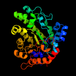
| 1 | c2infB_
|
|
 |
100.0 |
45 |
PDB header:lyase
Chain: B: PDB Molecule:uroporphyrinogen decarboxylase;
PDBTitle: crystal structure of uroporphyrinogen decarboxylase from bacillus2 subtilis
|
|
|
|
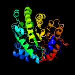
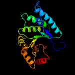
| 2 | c3cyvA_
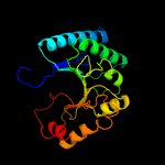
|
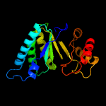
|
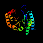
 |
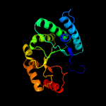
100.0 |
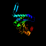
41 |
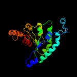
PDB header:lyase
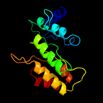
Chain: A: PDB Molecule:uroporphyrinogen decarboxylase;
PDBTitle: crystal structure of uroporphyrinogen decarboxylase from2 shigella flexineri: new insights into its catalytic3 mechanism
|
|
|
|
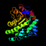
| 3 | d1r3sa_
|
|
 |
100.0 |
39 |
Fold:TIM beta/alpha-barrel
Superfamily:UROD/MetE-like
Family:Uroporphyrinogen decarboxylase, UROD |
|
|
|
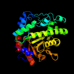
| 4 | c1jpkA_
|
|
 |
100.0 |
38 |
PDB header:lyase
Chain: A: PDB Molecule:uroporphyrinogen decarboxylase;
PDBTitle: gly156asp mutant of human urod, human uroporphyrinogen iii2 decarboxylase
|
|
|
|
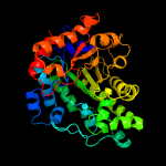
| 5 | c4exqA_
|
|
 |
100.0 |
43 |
PDB header:biosynthetic protein
Chain: A: PDB Molecule:uroporphyrinogen decarboxylase;
PDBTitle: crystal structure of uroporphyrinogen decarboxylase (upd) from2 burkholderia thailandensis e264
|
|
|
|
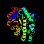
| 6 | d1j93a_
|
|
 |
100.0 |
37 |
Fold:TIM beta/alpha-barrel
Superfamily:UROD/MetE-like
Family:Uroporphyrinogen decarboxylase, UROD |
|
|
|
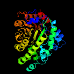
| 7 | c4zr8B_
|
|
 |
100.0 |
43 |
PDB header:lyase
Chain: B: PDB Molecule:uroporphyrinogen decarboxylase;
PDBTitle: structure of uroporphyrinogen decarboxylase from acinetobacter2 baumannii
|
|
|
|
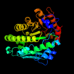
| 8 | c2ejaB_
|
|
 |
100.0 |
36 |
PDB header:lyase
Chain: B: PDB Molecule:uroporphyrinogen decarboxylase;
PDBTitle: crystal structure of uroporphyrinogen decarboxylase from2 aquifex aeolicus
|
|
|
|
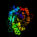
| 9 | c4ay8B_
|
|
 |
100.0 |
19 |
PDB header:transferase
Chain: B: PDB Molecule:methylcobalamin\: coenzyme m methyltransferase;
PDBTitle: semet-derivative of a methyltransferase from m. mazei
|
|
|
|
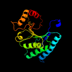
| 10 | d1u1ha2
|
|
 |
99.3 |
12 |
Fold:TIM beta/alpha-barrel
Superfamily:UROD/MetE-like
Family:Cobalamin-independent methionine synthase |
|
|
|
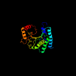
| 11 | c1u22A_
|
|
 |
99.1 |
12 |
PDB header:transferase
Chain: A: PDB Molecule:5-methyltetrahydropteroyltriglutamate--
PDBTitle: a. thaliana cobalamine independent methionine synthase
|
|
|
|
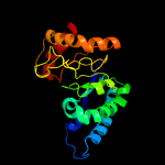
| 12 | c3rpdB_
|
|
 |
99.1 |
16 |
PDB header:transferase
Chain: B: PDB Molecule:methionine synthase (b12-independent);
PDBTitle: the structure of a b12-independent methionine synthase from shewanella2 sp. w3-18-1 in complex with selenomethionine.
|
|
|
|
| 13 | c4ztxA_
|
|
 |
99.1 |
15 |
PDB header:transferase
Chain: A: PDB Molecule:cobalamin-independent methionine synthase;
PDBTitle: neurospora crassa cobalamin-independent methionine synthase complexed2 with zn2+
|
|
|
|
| 14 | c3l7sA_
|
|
 |
99.1 |
16 |
PDB header:transferase
Chain: A: PDB Molecule:5-methyltetrahydropteroyltriglutamate--homocysteine
PDBTitle: crystal structure of mete coordinated with zinc from streptococcus2 mutans
|
|
|
|
| 15 | c2nq5A_
|
|
 |
99.0 |
17 |
PDB header:transferase
Chain: A: PDB Molecule:5-methyltetrahydropteroyltriglutamate--homocysteine
PDBTitle: crystal structure of methyltransferase from streptococcus mutans
|
|
|
|
| 16 | c3ppgA_
|
|
 |
99.0 |
15 |
PDB header:transferase
Chain: A: PDB Molecule:5-methyltetrahydropteroyltriglutamate--homocysteine
PDBTitle: crystal structure of the candida albicans methionine synthase by2 surface entropy reduction, alanine variant with zinc
|
|
|
|
| 17 | c1t7lA_
|
|
 |
99.0 |
13 |
PDB header:transferase
Chain: A: PDB Molecule:5-methyltetrahydropteroyltriglutamate--homocysteine
PDBTitle: crystal structure of cobalamin-independent methionine synthase from t.2 maritima
|
|
|
|
| 18 | c1ypxA_
|
|
 |
98.9 |
12 |
PDB header:transferase
Chain: A: PDB Molecule:putative vitamin-b12 independent methionine synthase family
PDBTitle: crystal structure of the putative vitamin-b12 independent methionine2 synthase from listeria monocytogenes, northeast structural genomics3 target lmr13
|
|
|
|
| 19 | d1u1ha1
|
|
 |
97.9 |
12 |
Fold:TIM beta/alpha-barrel
Superfamily:UROD/MetE-like
Family:Cobalamin-independent methionine synthase |
|
|
|
| 20 | c3lerA_
|
|
 |
96.7 |
14 |
PDB header:lyase
Chain: A: PDB Molecule:dihydrodipicolinate synthase;
PDBTitle: crystal structure of dihydrodipicolinate synthase from campylobacter2 jejuni subsp. jejuni nctc 11168
|
|
|
|
| 21 | c2h9aB_ |
|
not modelled |
96.7 |
11 |
PDB header:oxidoreductase
Chain: B: PDB Molecule:co dehydrogenase/acetyl-coa synthase, iron-sulfur protein;
PDBTitle: corrinoid iron-sulfur protein
|
|
|
| 22 | c5ud6B_ |
|
not modelled |
96.5 |
16 |
PDB header:lyase
Chain: B: PDB Molecule:dihydrodipicolinate synthase;
PDBTitle: crystal structure of dhdps from cyanidioschyzon merolae with lysine2 bound
|
|
|
| 23 | c2ehhE_ |
|
not modelled |
96.4 |
18 |
PDB header:lyase
Chain: E: PDB Molecule:dihydrodipicolinate synthase;
PDBTitle: crystal structure of dihydrodipicolinate synthase from2 aquifex aeolicus
|
|
|
| 24 | c3bolB_ |
|
not modelled |
96.3 |
18 |
PDB header:transferase
Chain: B: PDB Molecule:5-methyltetrahydrofolate s-homocysteine
PDBTitle: cobalamin-dependent methionine synthase (1-566) from2 thermotoga maritima complexed with zn2+
|
|
|
| 25 | c3daqB_ |
|
not modelled |
96.3 |
15 |
PDB header:lyase
Chain: B: PDB Molecule:dihydrodipicolinate synthase;
PDBTitle: crystal structure of dihydrodipicolinate synthase from methicillin-2 resistant staphylococcus aureus
|
|
|
| 26 | c4djdD_ |
|
not modelled |
96.2 |
14 |
PDB header:transferase/vitamin-binding protein
Chain: D: PDB Molecule:corrinoid/iron-sulfur protein small subunit;
PDBTitle: crystal structure of folate-free corrinoid iron-sulfur protein (cfesp)2 in complex with its methyltransferase (metr)
|
|
|
| 27 | c6mqhA_ |
|
not modelled |
96.1 |
20 |
PDB header:lyase
Chain: A: PDB Molecule:4-hydroxy-tetrahydrodipicolinate synthase;
PDBTitle: crystal structure of 4-hydroxy-tetrahydrodipicolinate synthase (htpa2 synthase) from burkholderia mallei
|
|
|
| 28 | c3lciA_ |
|
not modelled |
96.1 |
16 |
PDB header:lyase
Chain: A: PDB Molecule:n-acetylneuraminate lyase;
PDBTitle: the d-sialic acid aldolase mutant v251w
|
|
|
| 29 | d1xxxa1 |
|
not modelled |
96.1 |
14 |
Fold:TIM beta/alpha-barrel
Superfamily:Aldolase
Family:Class I aldolase |
|
|
| 30 | c3bi8A_ |
|
not modelled |
96.1 |
16 |
PDB header:lyase
Chain: A: PDB Molecule:dihydrodipicolinate synthase;
PDBTitle: structure of dihydrodipicolinate synthase from clostridium2 botulinum
|
|
|
| 31 | d1xkya1 |
|
not modelled |
96.0 |
15 |
Fold:TIM beta/alpha-barrel
Superfamily:Aldolase
Family:Class I aldolase |
|
|
| 32 | c2r8wB_ |
|
not modelled |
96.0 |
18 |
PDB header:lyase
Chain: B: PDB Molecule:agr_c_1641p;
PDBTitle: the crystal structure of dihydrodipicolinate synthase (atu0899) from2 agrobacterium tumefaciens str. c58
|
|
|
| 33 | c3d0cB_ |
|
not modelled |
96.0 |
15 |
PDB header:lyase
Chain: B: PDB Molecule:dihydrodipicolinate synthase;
PDBTitle: crystal structure of dihydrodipicolinate synthase from oceanobacillus2 iheyensis at 1.9 a resolution
|
|
|
| 34 | c4wuiA_ |
|
not modelled |
96.0 |
21 |
PDB header:isomerase
Chain: A: PDB Molecule:n-(5'-phosphoribosyl)anthranilate isomerase;
PDBTitle: crystal structure of trpf from jonesia denitrificans
|
|
|
| 35 | c3n2xB_ |
|
not modelled |
95.9 |
21 |
PDB header:lyase
Chain: B: PDB Molecule:uncharacterized protein yage;
PDBTitle: crystal structure of yage, a prophage protein belonging to the2 dihydrodipicolinic acid synthase family from e. coli k12 in complex3 with pyruvate
|
|
|
| 36 | c2rfgB_ |
|
not modelled |
95.9 |
14 |
PDB header:lyase
Chain: B: PDB Molecule:dihydrodipicolinate synthase;
PDBTitle: crystal structure of dihydrodipicolinate synthase from hahella2 chejuensis at 1.5a resolution
|
|
|
| 37 | c3noeA_ |
|
not modelled |
95.9 |
19 |
PDB header:lyase
Chain: A: PDB Molecule:dihydrodipicolinate synthase;
PDBTitle: crystal structure of dihydrodipicolinate synthase from pseudomonas2 aeruginosa
|
|
|
| 38 | c3pueA_ |
|
not modelled |
95.9 |
19 |
PDB header:lyase
Chain: A: PDB Molecule:dihydrodipicolinate synthase;
PDBTitle: crystal structure of the complex of dhydrodipicolinate synthase from2 acinetobacter baumannii with lysine at 2.6a resolution
|
|
|
| 39 | c4xkyC_ |
|
not modelled |
95.9 |
13 |
PDB header:lyase
Chain: C: PDB Molecule:dihydrodipicolinate synthase;
PDBTitle: structure of dihydrodipicolinate synthase from the commensal bacterium2 bacteroides thetaiotaomicron at 2.1 a resolution
|
|
|
| 40 | c6daoB_ |
|
not modelled |
95.8 |
24 |
PDB header:lyase
Chain: B: PDB Molecule:trans-o-hydroxybenzylidenepyruvate hydratase-aldolase;
PDBTitle: nahe wt selenomethionine
|
|
|
| 41 | c4ah7C_ |
|
not modelled |
95.8 |
12 |
PDB header:lyase
Chain: C: PDB Molecule:n-acetylneuraminate lyase;
PDBTitle: structure of wild type stapylococcus aureus n-acetylneuraminic acid2 lyase in complex with pyruvate
|
|
|
| 42 | c2v9dB_ |
|
not modelled |
95.8 |
21 |
PDB header:lyase
Chain: B: PDB Molecule:yage;
PDBTitle: crystal structure of yage, a prophage protein belonging to2 the dihydrodipicolinic acid synthase family from e. coli3 k12
|
|
|
| 43 | d1nsja_ |
|
not modelled |
95.8 |
18 |
Fold:TIM beta/alpha-barrel
Superfamily:Ribulose-phoshate binding barrel
Family:Tryptophan biosynthesis enzymes |
|
|
| 44 | c4nq1B_ |
|
not modelled |
95.7 |
19 |
PDB header:lyase
Chain: B: PDB Molecule:4-hydroxy-tetrahydrodipicolinate synthase;
PDBTitle: legionella pneumophila dihydrodipicolinate synthase with first2 substrate pyruvate bound in the active site
|
|
|
| 45 | c3h5dD_ |
|
not modelled |
95.7 |
17 |
PDB header:lyase
Chain: D: PDB Molecule:dihydrodipicolinate synthase;
PDBTitle: dihydrodipicolinate synthase from drug-resistant streptococcus2 pneumoniae
|
|
|
| 46 | d1hl2a_ |
|
not modelled |
95.6 |
17 |
Fold:TIM beta/alpha-barrel
Superfamily:Aldolase
Family:Class I aldolase |
|
|
| 47 | c1piiA_ |
|
not modelled |
95.5 |
16 |
PDB header:bifunctional(isomerase and synthase)
Chain: A: PDB Molecule:n-(5'phosphoribosyl)anthranilate isomerase;
PDBTitle: three-dimensional structure of the bifunctional enzyme2 phosphoribosylanthranilate isomerase: indoleglycerolphosphate3 synthase from escherichia coli refined at 2.0 angstroms resolution
|
|
|
| 48 | c3cprB_ |
|
not modelled |
95.4 |
13 |
PDB header:lyase
Chain: B: PDB Molecule:dihydrodipicolinate synthetase;
PDBTitle: the crystal structure of corynebacterium glutamicum2 dihydrodipicolinate synthase to 2.2 a resolution
|
|
|
| 49 | c2r94B_ |
|
not modelled |
95.4 |
16 |
PDB header:lyase
Chain: B: PDB Molecule:2-keto-3-deoxy-(6-phospho-)gluconate aldolase;
PDBTitle: crystal structure of kd(p)ga from t.tenax
|
|
|
| 50 | c6daqA_ |
|
not modelled |
95.3 |
14 |
PDB header:lyase
Chain: A: PDB Molecule:phdj;
PDBTitle: phdj bound to substrate intermediate
|
|
|
| 51 | c3dz1A_ |
|
not modelled |
95.3 |
14 |
PDB header:lyase
Chain: A: PDB Molecule:dihydrodipicolinate synthase;
PDBTitle: crystal structure of dihydrodipicolinate synthase from2 rhodopseudomonas palustris at 1.87a resolution
|
|
|
| 52 | d1o5ka_ |
|
not modelled |
95.2 |
15 |
Fold:TIM beta/alpha-barrel
Superfamily:Aldolase
Family:Class I aldolase |
|
|
| 53 | d2a6na1 |
|
not modelled |
95.2 |
15 |
Fold:TIM beta/alpha-barrel
Superfamily:Aldolase
Family:Class I aldolase |
|
|
| 54 | c3na8A_ |
|
not modelled |
95.2 |
17 |
PDB header:lyase
Chain: A: PDB Molecule:putative dihydrodipicolinate synthetase;
PDBTitle: crystal structure of a putative dihydrodipicolinate synthetase from2 pseudomonas aeruginosa
|
|
|
| 55 | d1w3ia_ |
|
not modelled |
95.2 |
16 |
Fold:TIM beta/alpha-barrel
Superfamily:Aldolase
Family:Class I aldolase |
|
|
| 56 | c2yxgD_ |
|
not modelled |
95.0 |
14 |
PDB header:lyase
Chain: D: PDB Molecule:dihydrodipicolinate synthase;
PDBTitle: crystal structure of dihyrodipicolinate synthase (dapa)
|
|
|
| 57 | c2vc6A_ |
|
not modelled |
94.9 |
15 |
PDB header:lyase
Chain: A: PDB Molecule:dihydrodipicolinate synthase;
PDBTitle: structure of mosa from s. meliloti with pyruvate bound
|
|
|
| 58 | c2hmcA_ |
|
not modelled |
94.9 |
20 |
PDB header:structural genomics, unknown function
Chain: A: PDB Molecule:dihydrodipicolinate synthase;
PDBTitle: the crystal structure of dihydrodipicolinate synthase dapa from2 agrobacterium tumefaciens
|
|
|
| 59 | c5ui3C_ |
|
not modelled |
94.9 |
17 |
PDB header:lyase
Chain: C: PDB Molecule:dihydrodipicolinate synthase;
PDBTitle: crystal structure of dhdps from chlamydomonas reinhardtii
|
|
|
| 60 | c5visB_ |
|
not modelled |
94.8 |
16 |
PDB header:hydrolase,oxidoreductase
Chain: B: PDB Molecule:dihydropteroate synthase;
PDBTitle: 1.73 angstrom resolution crystal structure of dihydropteroate synthase2 (folp-smz_b27) from soil uncultured bacterium.
|
|
|
| 61 | d1ad1a_ |
|
not modelled |
94.8 |
11 |
Fold:TIM beta/alpha-barrel
Superfamily:Dihydropteroate synthetase-like
Family:Dihydropteroate synthetase |
|
|
| 62 | c6h4eB_ |
|
not modelled |
94.8 |
14 |
PDB header:lyase
Chain: B: PDB Molecule:putative n-acetylneuraminate lyase;
PDBTitle: proteus mirabilis n-acetylneuraminate lyase
|
|
|
| 63 | c5ey5A_ |
|
not modelled |
94.8 |
18 |
PDB header:lyase
Chain: A: PDB Molecule:lbcats-a;
PDBTitle: lbcats
|
|
|
| 64 | c5ktlA_ |
|
not modelled |
94.8 |
15 |
PDB header:lyase
Chain: A: PDB Molecule:4-hydroxy-tetrahydrodipicolinate synthase;
PDBTitle: dihydrodipicolinate synthase from the industrial and evolutionarily2 important cyanobacteria anabaena variabilis.
|
|
|
| 65 | c5afdA_ |
|
not modelled |
94.7 |
17 |
PDB header:lyase
Chain: A: PDB Molecule:n-acetylneuraminate lyase;
PDBTitle: native structure of n-acetylneuramininate lyase (sialic acid aldolase)2 from aliivibrio salmonicida
|
|
|
| 66 | c6arhA_ |
|
not modelled |
94.6 |
18 |
PDB header:lyase
Chain: A: PDB Molecule:n-acetylneuraminate lyase;
PDBTitle: crystal structure of human nal at a resolution of 1.6 angstrom
|
|
|
| 67 | c4n4qD_ |
|
not modelled |
94.6 |
12 |
PDB header:lyase
Chain: D: PDB Molecule:acylneuraminate lyase;
PDBTitle: crystal structure of n-acetylneuraminate lyase from mycoplasma2 synoviae, crystal form ii
|
|
|
| 68 | c5c54D_ |
|
not modelled |
94.6 |
18 |
PDB header:lyase
Chain: D: PDB Molecule:dihydrodipicolinate synthase/n-acetylneuraminate lyase;
PDBTitle: crystal structure of a novel n-acetylneuraminic acid lyase from2 corynebacterium glutamicum
|
|
|
| 69 | c4i7vD_ |
|
not modelled |
94.5 |
17 |
PDB header:biosynthetic protein
Chain: D: PDB Molecule:dihydrodipicolinate synthase;
PDBTitle: agrobacterium tumefaciens dhdps with pyruvate
|
|
|
| 70 | c3fluD_ |
|
not modelled |
94.5 |
17 |
PDB header:lyase
Chain: D: PDB Molecule:dihydrodipicolinate synthase;
PDBTitle: crystal structure of dihydrodipicolinate synthase from the pathogen2 neisseria meningitidis
|
|
|
| 71 | c3s5oA_ |
|
not modelled |
94.5 |
13 |
PDB header:lyase
Chain: A: PDB Molecule:4-hydroxy-2-oxoglutarate aldolase, mitochondrial;
PDBTitle: crystal structure of human 4-hydroxy-2-oxoglutarate aldolase bound to2 pyruvate
|
|
|
| 72 | c3g0sA_ |
|
not modelled |
94.5 |
16 |
PDB header:lyase
Chain: A: PDB Molecule:dihydrodipicolinate synthase;
PDBTitle: dihydrodipicolinate synthase from salmonella typhimurium lt2
|
|
|
| 73 | c4dppB_ |
|
not modelled |
94.5 |
15 |
PDB header:lyase
Chain: B: PDB Molecule:dihydrodipicolinate synthase 2, chloroplastic;
PDBTitle: the structure of dihydrodipicolinate synthase 2 from arabidopsis2 thaliana
|
|
|
| 74 | c5ks8F_ |
|
not modelled |
94.4 |
23 |
PDB header:ligase
Chain: F: PDB Molecule:pyruvate carboxylase subunit beta;
PDBTitle: crystal structure of two-subunit pyruvate carboxylase from2 methylobacillus flagellatus
|
|
|
| 75 | c3e96B_ |
|
not modelled |
94.4 |
16 |
PDB header:lyase
Chain: B: PDB Molecule:dihydrodipicolinate synthase;
PDBTitle: crystal structure of dihydrodipicolinate synthase from bacillus2 clausii
|
|
|
| 76 | c2nuxB_ |
|
not modelled |
94.4 |
16 |
PDB header:lyase
Chain: B: PDB Molecule:2-keto-3-deoxygluconate/2-keto-3-deoxy-6-phospho gluconate
PDBTitle: 2-keto-3-deoxygluconate aldolase from sulfolobus acidocaldarius,2 native structure in p6522 at 2.5 a resolution
|
|
|
| 77 | c3bg3B_ |
|
not modelled |
94.4 |
16 |
PDB header:ligase
Chain: B: PDB Molecule:pyruvate carboxylase, mitochondrial;
PDBTitle: crystal structure of human pyruvate carboxylase (missing the biotin2 carboxylase domain at the n-terminus)
|
|
|
| 78 | c3qfeB_ |
|
not modelled |
94.3 |
12 |
PDB header:lyase
Chain: B: PDB Molecule:putative dihydrodipicolinate synthase family protein;
PDBTitle: crystal structures of a putative dihydrodipicolinate synthase family2 protein from coccidioides immitis
|
|
|
| 79 | c4qslE_ |
|
not modelled |
94.3 |
23 |
PDB header:ligase
Chain: E: PDB Molecule:pyruvate carboxylase;
PDBTitle: crystal structure of listeria monocytogenes pyruvate carboxylase
|
|
|
| 80 | c3si9B_ |
|
not modelled |
94.3 |
12 |
PDB header:lyase
Chain: B: PDB Molecule:dihydrodipicolinate synthase;
PDBTitle: crystal structure of dihydrodipicolinate synthase from bartonella2 henselae
|
|
|
| 81 | c3bg5C_ |
|
not modelled |
94.2 |
16 |
PDB header:ligase
Chain: C: PDB Molecule:pyruvate carboxylase;
PDBTitle: crystal structure of staphylococcus aureus pyruvate carboxylase
|
|
|
| 82 | d1f74a_ |
|
not modelled |
94.2 |
15 |
Fold:TIM beta/alpha-barrel
Superfamily:Aldolase
Family:Class I aldolase |
|
|
| 83 | c6omzA_ |
|
not modelled |
94.2 |
18 |
PDB header:ligase
Chain: A: PDB Molecule:dihydropteroate synthase;
PDBTitle: crystal structure of dihydropteroate synthase from mycobacterium2 smegmatis with bound 6-hydroxymethylpterin-monophosphate
|
|
|
| 84 | c3eb2A_ |
|
not modelled |
94.1 |
20 |
PDB header:lyase
Chain: A: PDB Molecule:putative dihydrodipicolinate synthetase;
PDBTitle: crystal structure of dihydrodipicolinate synthase from2 rhodopseudomonas palustris at 2.0a resolution
|
|
|
| 85 | c4qslC_ |
|
not modelled |
94.0 |
18 |
PDB header:ligase
Chain: C: PDB Molecule:pyruvate carboxylase;
PDBTitle: crystal structure of listeria monocytogenes pyruvate carboxylase
|
|
|
| 86 | c4icnB_ |
|
not modelled |
93.9 |
21 |
PDB header:lyase
Chain: B: PDB Molecule:dihydrodipicolinate synthase;
PDBTitle: dihydrodipicolinate synthase from shewanella benthica
|
|
|
| 87 | c3f4cA_ |
|
not modelled |
93.9 |
11 |
PDB header:hydrolase
Chain: A: PDB Molecule:organophosphorus hydrolase;
PDBTitle: crystal structure of organophosphorus hydrolase from geobacillus2 stearothermophilus strain 10, with glycerol bound
|
|
|
| 88 | c3tn6A_ |
|
not modelled |
93.6 |
11 |
PDB header:hydrolase
Chain: A: PDB Molecule:phosphotriesterase;
PDBTitle: crystal structure of gkap mutant r230h from geobacillus kaustophilus2 hta426
|
|
|
| 89 | c3b4uB_ |
|
not modelled |
93.6 |
13 |
PDB header:lyase
Chain: B: PDB Molecule:dihydrodipicolinate synthase;
PDBTitle: crystal structure of dihydrodipicolinate synthase from agrobacterium2 tumefaciens str. c58
|
|
|
| 90 | c1rr2A_ |
|
not modelled |
93.5 |
15 |
PDB header:transferase
Chain: A: PDB Molecule:transcarboxylase 5s subunit;
PDBTitle: propionibacterium shermanii transcarboxylase 5s subunit bound to 2-2 ketobutyric acid
|
|
|
| 91 | c4ur7B_ |
|
not modelled |
93.5 |
12 |
PDB header:lyase
Chain: B: PDB Molecule:keto-deoxy-d-galactarate dehydratase;
PDBTitle: crystal structure of keto-deoxy-d-galactarate dehydratase2 complexed with pyruvate
|
|
|
| 92 | c5ks8D_ |
|
not modelled |
93.5 |
25 |
PDB header:ligase
Chain: D: PDB Molecule:pyruvate carboxylase subunit beta;
PDBTitle: crystal structure of two-subunit pyruvate carboxylase from2 methylobacillus flagellatus
|
|
|
| 93 | c1tx2A_ |
|
not modelled |
93.4 |
16 |
PDB header:transferase
Chain: A: PDB Molecule:dhps, dihydropteroate synthase;
PDBTitle: dihydropteroate synthetase, with bound inhibitor manic, from bacillus2 anthracis
|
|
|
| 94 | d1tx2a_ |
|
not modelled |
93.4 |
16 |
Fold:TIM beta/alpha-barrel
Superfamily:Dihydropteroate synthetase-like
Family:Dihydropteroate synthetase |
|
|
| 95 | c3pnzD_ |
|
not modelled |
93.4 |
12 |
PDB header:hydrolase
Chain: D: PDB Molecule:phosphotriesterase family protein;
PDBTitle: crystal structure of the lactonase lmo2620 from listeria monocytogenes
|
|
|
| 96 | c4uxdC_ |
|
not modelled |
93.2 |
11 |
PDB header:lyase
Chain: C: PDB Molecule:2-dehydro-3-deoxy-d-gluconate/2-dehydro-3-deoxy-
PDBTitle: 2-keto 3-deoxygluconate aldolase from picrophilus torridus
|
|
|
| 97 | c6qkgB_ |
|
not modelled |
93.1 |
12 |
PDB header:flavoprotein
Chain: B: PDB Molecule:ncr a;
PDBTitle: 2-naphthoyl-coa reductase(ncr)
|
|
|
| 98 | c2nx9B_ |
|
not modelled |
93.1 |
25 |
PDB header:lyase
Chain: B: PDB Molecule:oxaloacetate decarboxylase 2, subunit alpha;
PDBTitle: crystal structure of the carboxyltransferase domain of the2 oxaloacetate decarboxylase na+ pump from vibrio cholerae
|
|
|
| 99 | d1piia1 |
|
not modelled |
93.0 |
17 |
Fold:TIM beta/alpha-barrel
Superfamily:Ribulose-phoshate binding barrel
Family:Tryptophan biosynthesis enzymes |
|
|
| 100 | c3bg3A_ |
|
not modelled |
92.7 |
19 |
PDB header:ligase
Chain: A: PDB Molecule:pyruvate carboxylase, mitochondrial;
PDBTitle: crystal structure of human pyruvate carboxylase (missing the biotin2 carboxylase domain at the n-terminus)
|
|
|
| 101 | c2y5sA_ |
|
not modelled |
92.7 |
18 |
PDB header:transferase
Chain: A: PDB Molecule:dihydropteroate synthase;
PDBTitle: crystal structure of burkholderia cenocepacia dihydropteroate2 synthase complexed with 7,8-dihydropteroate.
|
|
|
| 102 | c4qskB_ |
|
not modelled |
92.7 |
21 |
PDB header:ligase
Chain: B: PDB Molecule:pyruvate carboxylase;
PDBTitle: crystal structure of l. monocytogenes pyruvate carboxylase in complex2 with cyclic-di-amp
|
|
|
| 103 | c4o1fB_ |
|
not modelled |
92.5 |
14 |
PDB header:transferase
Chain: B: PDB Molecule:dihydropteroate synthase dhps;
PDBTitle: structure of a methyltransferase component in complex with thf2 involved in o-demethylation
|
|
|
| 104 | c2vc7A_ |
|
not modelled |
92.2 |
11 |
PDB header:hydrolase
Chain: A: PDB Molecule:aryldialkylphosphatase;
PDBTitle: structural basis for natural lactonase and promiscuous2 phosphotriesterase activities
|
|
|
| 105 | c3hf3A_ |
|
not modelled |
92.0 |
22 |
PDB header:oxidoreductase
Chain: A: PDB Molecule:chromate reductase;
PDBTitle: old yellow enzyme from thermus scotoductus sa-01
|
|
|
| 106 | c3bleA_ |
|
not modelled |
92.0 |
18 |
PDB header:transferase
Chain: A: PDB Molecule:citramalate synthase from leptospira interrogans;
PDBTitle: crystal structure of the catalytic domain of licms in complexed with2 malonate
|
|
|
| 107 | c3k30B_ |
|
not modelled |
91.8 |
15 |
PDB header:oxidoreductase
Chain: B: PDB Molecule:histamine dehydrogenase;
PDBTitle: histamine dehydrogenase from nocardiodes simplex
|
|
|
| 108 | c2yciX_ |
|
not modelled |
91.8 |
8 |
PDB header:transferase
Chain: X: PDB Molecule:5-methyltetrahydrofolate corrinoid/iron sulfur protein
PDBTitle: methyltransferase native
|
|
|
| 109 | c4lrtC_ |
|
not modelled |
91.8 |
25 |
PDB header:lyase/oxidoreductase
Chain: C: PDB Molecule:4-hydroxy-2-oxovalerate aldolase;
PDBTitle: crystal and solution structures of the bifunctional enzyme2 (aldolase/aldehyde dehydrogenase) from thermomonospora curvata,3 reveal a cofactor-binding domain motion during nad+ and coa4 accommodation whithin the shared cofactor-binding site
|
|
|
| 110 | c2cw6B_ |
|
not modelled |
91.8 |
13 |
PDB header:lyase
Chain: B: PDB Molecule:hydroxymethylglutaryl-coa lyase, mitochondrial;
PDBTitle: crystal structure of human hmg-coa lyase: insights into2 catalysis and the molecular basis for3 hydroxymethylglutaric aciduria
|
|
|
| 111 | c3gndC_ |
|
not modelled |
91.7 |
13 |
PDB header:lyase
Chain: C: PDB Molecule:aldolase lsrf;
PDBTitle: crystal structure of e. coli lsrf in complex with ribulose-5-phosphate
|
|
|
| 112 | c1nvmG_ |
|
not modelled |
91.7 |
20 |
PDB header:lyase/oxidoreductase
Chain: G: PDB Molecule:4-hydroxy-2-oxovalerate aldolase;
PDBTitle: crystal structure of a bifunctional aldolase-dehydrogenase :2 sequestering a reactive and volatile intermediate
|
|
|
| 113 | c6cluC_ |
|
not modelled |
91.6 |
15 |
PDB header:antimicrobial protein
Chain: C: PDB Molecule:dihydropteroate synthase;
PDBTitle: staphylococcus aureus dihydropteroate synthase (sadhps) f17l e208k2 double mutant structure
|
|
|
| 114 | d1vlia2 |
|
not modelled |
91.6 |
16 |
Fold:TIM beta/alpha-barrel
Superfamily:Aldolase
Family:NeuB-like |
|
|
| 115 | d3bofa1 |
|
not modelled |
91.6 |
10 |
Fold:TIM beta/alpha-barrel
Superfamily:Dihydropteroate synthetase-like
Family:Methyltetrahydrofolate-utiluzing methyltransferases |
|
|
| 116 | c1zcoA_ |
|
not modelled |
91.5 |
12 |
PDB header:lyase
Chain: A: PDB Molecule:2-dehydro-3-deoxyphosphoheptonate aldolase;
PDBTitle: crystal structure of pyrococcus furiosus 3-deoxy-d-arabino-2 heptulosonate 7-phosphate synthase
|
|
|
| 117 | c4jn6C_ |
|
not modelled |
91.4 |
27 |
PDB header:lyase/oxidoreductase
Chain: C: PDB Molecule:4-hydroxy-2-oxovalerate aldolase;
PDBTitle: crystal structure of the aldolase-dehydrogenase complex from2 mycobacterium tuberculosis hrv37
|
|
|
| 118 | c4tmcB_ |
|
not modelled |
91.4 |
17 |
PDB header:flavoprotein
Chain: B: PDB Molecule:old yellow enzyme;
PDBTitle: crystal structure of old yellow enzyme from candida macedoniensis2 aku4588 complexed with p-hydroxybenzaldehyde
|
|
|
| 119 | c4aajA_ |
|
not modelled |
91.2 |
14 |
PDB header:isomerase
Chain: A: PDB Molecule:n-(5'-phosphoribosyl)anthranilate isomerase;
PDBTitle: structure of n-(5'-phosphoribosyl)anthranilate isomerase from2 pyrococcus furiosus
|
|
|
| 120 | c1ydnA_ |
|
not modelled |
91.2 |
15 |
PDB header:lyase
Chain: A: PDB Molecule:hydroxymethylglutaryl-coa lyase;
PDBTitle: crystal structure of the hmg-coa lyase from brucella melitensis,2 northeast structural genomics target lr35.
|
|
|


