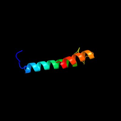
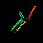
1 c5fmnB_
47.3
17
PDB header: dna binding proteinChain: B: PDB Molecule: inrs;PDBTitle: the nickel-responsive transcriptional regulator inrs
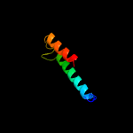
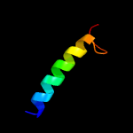
2 c5lbmD_
41.1
23
PDB header: transcriptionChain: D: PDB Molecule: transcriptional repressor frmr;PDBTitle: the asymmetric tetrameric structure of the formaldehyde sensing2 transcriptional repressor frmr from escherichia coli
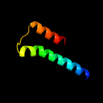
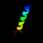
3 c4m1pA_
40.1
17
PDB header: transcription repressorChain: A: PDB Molecule: copper-sensitive operon repressor (csor);PDBTitle: crystal structure of the copper-sensing repressor csor with cu(i) from2 geobacillus thermodenitrificans ng80-2
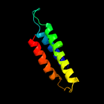
4 c6e8wC_
39.1
19
PDB header: viral proteinChain: C: PDB Molecule: envelope glycoprotein gp160;PDBTitle: mper-tm domain of hiv-1 envelope glycoprotein (env)
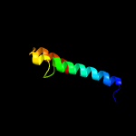
5 c5lcyD_
37.3
17
PDB header: transcriptionChain: D: PDB Molecule: frmr;PDBTitle: formaldehyde-responsive regulator frmr e64h variant from salmonella2 enterica serovar typhimurium
6 c2f49C_
32.5
47
PDB header: transferaseChain: C: PDB Molecule: ste5 peptide;PDBTitle: crystal structure of fus3 in complex with a ste5 peptide
7 c5yc0Q_
25.4
21
PDB header: viral protein/inhibitorChain: Q: PDB Molecule: lp-46;PDBTitle: crystal structure of lp-46/n44
8 c5yc0W_
25.4
21
PDB header: viral protein/inhibitorChain: W: PDB Molecule: lp-46;PDBTitle: crystal structure of lp-46/n44
9 c5yc0I_
25.4
21
PDB header: viral protein/inhibitorChain: I: PDB Molecule: lp-46;PDBTitle: crystal structure of lp-46/n44
10 c5yc0P_
24.3
21
PDB header: viral protein/inhibitorChain: P: PDB Molecule: lp-46;PDBTitle: crystal structure of lp-46/n44
11 c5yc0G_
24.3
21
PDB header: viral protein/inhibitorChain: G: PDB Molecule: lp-46;PDBTitle: crystal structure of lp-46/n44
12 c2sivD_
21.3
21
PDB header: envelope glycoproteinChain: D: PDB Molecule: siv gp41 glycoprotein;PDBTitle: siv gp41 core structure
13 c5yqrA_
19.7
26
PDB header: transport proteinChain: A: PDB Molecule: endolysin/membrane-anchored lipid-binding protein lam6PDBTitle: crystal structure of the ph-like domain of lam6
14 c6btmD_
19.6
15
PDB header: membrane proteinChain: D: PDB Molecule: alternative complex iii subunit d;PDBTitle: structure of alternative complex iii from flavobacterium johnsoniae2 (wild type)
15 c5jr0A_
17.7
23
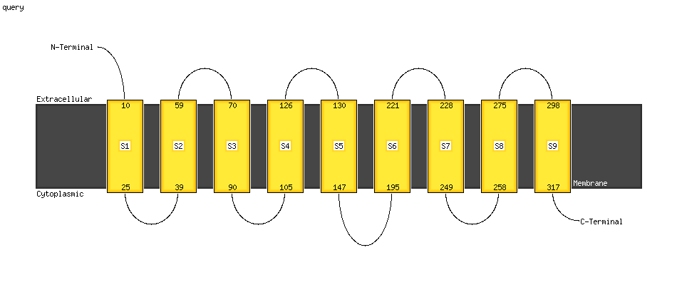
PDB header: metal binding proteinChain: A: PDB Molecule: sodium channel protein type 4 subunit alpha;PDBTitle: domain 4 segment 6 of voltage-gated sodium channel nav1.4
16 c2bg9B_
17.6
16
PDB header: ion channel/receptorChain: B: PDB Molecule: acetylcholine receptor protein, beta chain;PDBTitle: refined structure of the nicotinic acetylcholine receptor2 at 4a resolution.
17 c1yewI_
17.4
9
PDB header: oxidoreductase, membrane proteinChain: I: PDB Molecule: particulate methane monooxygenase, b subunit;PDBTitle: crystal structure of particulate methane monooxygenase
18 c3rgbA_
17.4
9
PDB header: oxidoreductaseChain: A: PDB Molecule: methane monooxygenase subunit b2;PDBTitle: crystal structure of particulate methane monooxygenase from2 methylococcus capsulatus (bath)
19 c6dmoA_
17.4
11
PDB header: protein bindingChain: A: PDB Molecule: protein patched homolog 1;PDBTitle: cryo-em structure of human ptch1 with three mutations2 l282q/t500f/p504l
20 c4adzA_
16.7
20
PDB header: transcriptionChain: A: PDB Molecule: csor;PDBTitle: crystal structure of the apo form of a copper-sensitive operon2 regulator (csor) protein from streptomyces lividans
21 c3h00A_
not modelled
16.0
25
PDB header: viral proteinChain: A: PDB Molecule: envelope glycoprotein gp160;PDBTitle: structure of the c-terminal domain of a putative hiv-1 gp41 fusion2 intermediate
22 c5yc0H_
not modelled
14.8
17
PDB header: viral protein/inhibitorChain: H: PDB Molecule: lp-46;PDBTitle: crystal structure of lp-46/n44
23 c2vl0A_
not modelled
14.3
13
PDB header: membrane proteinChain: A: PDB Molecule: elic pentameric ligand gated ion channel fromPDBTitle: x-ray structure of a pentameric ligand gated ion channel2 from erwinia chrysanthemi (elic)
24 c4qidB_
not modelled
14.1
10
PDB header: membrane proteinChain: B: PDB Molecule: bacteriorhodopsin-i;PDBTitle: crystal structure of haloquadratum walsbyi bacteriorhodopsin
25 c5wdaL_
not modelled
13.9
19
PDB header: protein transportChain: L: PDB Molecule: general secretion pathway protein g;PDBTitle: structure of the pulg pseudopilus
26 c3u3iA_
not modelled
13.4
23
PDB header: rna binding proteinChain: A: PDB Molecule: nucleocapsid protein;PDBTitle: a rna binding protein from crimean-congo hemorrhagic fever virus
27 c6ckoC_
not modelled
13.2
22
PDB header: dna binding protein/transferaseChain: C: PDB Molecule: histone-lysine n-methyltransferase, h3 lysine-79 specific;PDBTitle: crystal structure of an af10 fragment
28 c6ckoD_
not modelled
13.2
22
PDB header: dna binding protein/transferaseChain: D: PDB Molecule: histone-lysine n-methyltransferase, h3 lysine-79 specific;PDBTitle: crystal structure of an af10 fragment
29 c6nr2A_
not modelled
12.8
22
PDB header: transport proteinChain: A: PDB Molecule: transient receptor potential cation channel subfamily mPDBTitle: cryo-em structure of the trpm8 ion channel in complex with the menthol2 analog ws-12 and pi(4,5)p2
30 c6nr2C_
not modelled
12.8
22
PDB header: transport proteinChain: C: PDB Molecule: transient receptor potential cation channel subfamily mPDBTitle: cryo-em structure of the trpm8 ion channel in complex with the menthol2 analog ws-12 and pi(4,5)p2
31 c6nr2B_
not modelled
12.8
22
PDB header: transport proteinChain: B: PDB Molecule: transient receptor potential cation channel subfamily mPDBTitle: cryo-em structure of the trpm8 ion channel in complex with the menthol2 analog ws-12 and pi(4,5)p2
32 c6nr2D_
not modelled
12.8
22
PDB header: transport proteinChain: D: PDB Molecule: transient receptor potential cation channel subfamily mPDBTitle: cryo-em structure of the trpm8 ion channel in complex with the menthol2 analog ws-12 and pi(4,5)p2
33 c3k07A_
not modelled
12.5
14
PDB header: transport proteinChain: A: PDB Molecule: cation efflux system protein cusa;PDBTitle: crystal structure of cusa
34 c4aq5B_
not modelled
12.4
16
PDB header: membrane proteinChain: B: PDB Molecule: acetylcholine receptor beta subunit;PDBTitle: gating movement in acetylcholine receptor analysed by time-resolved2 electron cryo-microscopy (closed class)
35 c3wt9A_
not modelled
11.9
15
PDB header: proton transportChain: A: PDB Molecule: rhodopsin i;PDBTitle: crystal structure of the cell-free synthesized membrane protein,2 acetabularia rhodopsin i, at 1.48 angstrom
36 d1h2sa_
not modelled
11.8
17
Fold: Family A G protein-coupled receptor-likeSuperfamily: Family A G protein-coupled receptor-likeFamily: Bacteriorhodopsin-like
37 c4pj0L_
not modelled
11.6
22
PDB header: oxidoreductase, electron transportChain: L: PDB Molecule: photosystem ii reaction center protein l;PDBTitle: structure of t.elongatus photosystem ii, rows of dimers crystal2 packing
38 c4pj0l_
not modelled
11.6
22
PDB header: oxidoreductase, electron transportChain: L: PDB Molecule: photosystem ii reaction center protein l;PDBTitle: structure of t.elongatus photosystem ii, rows of dimers crystal2 packing
39 c4ub6l_
not modelled
11.4
22
PDB header: electron transport, photosynthesisChain: L: PDB Molecule: photosystem ii reaction center protein l;PDBTitle: native structure of photosystem ii (dataset-1) by a femtosecond x-ray2 laser
40 c5e7cl_
not modelled
11.4
22
PDB header: photosynthesisChain: L: PDB Molecule: photosystem ii reaction center protein l;PDBTitle: macromolecular diffractive imaging using imperfect crystals - bragg2 data
41 c4ixql_
not modelled
11.4
22
PDB header: photosynthesisChain: L: PDB Molecule: photosystem ii reaction center protein l;PDBTitle: rt fs x-ray diffraction of photosystem ii, dark state
42 c4fbyd_
not modelled
11.4
22
PDB header: photosynthesisChain: D: PDB Molecule: photosystem ii d2 protein;PDBTitle: fs x-ray diffraction of photosystem ii
43 c3bz2L_
not modelled
11.4
22
PDB header: electron transportChain: L: PDB Molecule: photosystem ii reaction center protein l;PDBTitle: crystal structure of cyanobacterial photosystem ii (part 2 of 2). this2 file contains second monomer of psii dimer
44 c4ub8l_
not modelled
11.4
22
PDB header: electron transport, photosynthesisChain: L: PDB Molecule: photosystem ii reaction center protein l;PDBTitle: native structure of photosystem ii (dataset-2) by a femtosecond x-ray2 laser
45 c1s5lL_
not modelled
11.4
22
PDB header: photosynthesisChain: L: PDB Molecule: photosystem ii reaction center l protein;PDBTitle: architecture of the photosynthetic oxygen evolving center
46 c4fbyL_
not modelled
11.4
22
PDB header: photosynthesisChain: L: PDB Molecule: photosystem ii reaction center protein l;PDBTitle: fs x-ray diffraction of photosystem ii
47 c4tniL_
not modelled
11.4
22
PDB header: electron transport,photosynthesisChain: L: PDB Molecule: photosystem ii reaction center protein l;PDBTitle: rt xfel structure of photosystem ii 500 ms after the third2 illumination at 4.6 a resolution
48 c4il6l_
not modelled
11.4
22
PDB header: electron transportChain: L: PDB Molecule: photosystem ii reaction center protein l;PDBTitle: structure of sr-substituted photosystem ii
49 c2axtl_
not modelled
11.4
22
PDB header: electron transportChain: L: PDB Molecule: photosystem ii reaction center l protein;PDBTitle: crystal structure of photosystem ii from thermosynechococcus elongatus
50 c4rvyL_
not modelled
11.4
22
PDB header: oxidoreductaseChain: L: PDB Molecule: photosystem ii reaction center protein l;PDBTitle: serial time resolved crystallography of photosystem ii using a2 femtosecond x-ray laser. the s state after two flashes (s3)
51 c4rvyl_
not modelled
11.4
22
PDB header: oxidoreductaseChain: L: PDB Molecule: photosystem ii reaction center protein l;PDBTitle: serial time resolved crystallography of photosystem ii using a2 femtosecond x-ray laser. the s state after two flashes (s3)
52 c4tnhL_
not modelled
11.4
22
PDB header: electron transport,photosynthesisChain: L: PDB Molecule: photosystem ii reaction center protein l;PDBTitle: rt xfel structure of photosystem ii in the dark state at 4.9 a2 resolution
53 c3prqL_
not modelled
11.4
22
PDB header: photosynthesisChain: L: PDB Molecule: photosystem ii reaction center protein l;PDBTitle: crystal structure of cyanobacterial photosystem ii in complex with2 terbutryn (part 1 of 2). this file contains first monomer of psii3 dimer
54 c5e7cL_
not modelled
11.4
22
PDB header: photosynthesisChain: L: PDB Molecule: photosystem ii reaction center protein l;PDBTitle: macromolecular diffractive imaging using imperfect crystals - bragg2 data
55 c3kziL_
not modelled
11.4
22
PDB header: electron transportChain: L: PDB Molecule: photosystem ii reaction center protein l;PDBTitle: crystal structure of monomeric form of cyanobacterial photosystem ii
56 c3arcL_
not modelled
11.4
22
PDB header: electron transport, photosynthesisChain: L: PDB Molecule: photosystem ii reaction center protein l;PDBTitle: crystal structure of oxygen-evolving photosystem ii at 1.9 angstrom2 resolution
57 c3bz1L_
not modelled
11.4
22
PDB header: electron transportChain: L: PDB Molecule: photosystem ii reaction center protein l;PDBTitle: crystal structure of cyanobacterial photosystem ii (part 1 of 2). this2 file contains first monomer of psii dimer
58 c4ixrl_
not modelled
11.4
22
PDB header: photosynthesisChain: L: PDB Molecule: photosystem ii reaction center protein l;PDBTitle: rt fs x-ray diffraction of photosystem ii, first illuminated state
59 c2axtL_
not modelled
11.4
22
PDB header: electron transportChain: L: PDB Molecule: photosystem ii reaction center l protein;PDBTitle: crystal structure of photosystem ii from thermosynechococcus elongatus
60 d2axtl1
not modelled
11.4
22
Fold: Single transmembrane helixSuperfamily: Photosystem II reaction center protein L, PsbLFamily: PsbL-like
61 c4tnkl_
not modelled
11.4
22
PDB header: electron transport,photosynthesisChain: L: PDB Molecule: photosystem ii reaction center protein l;PDBTitle: rt xfel structure of photosystem ii 250 microsec after the third2 illumination at 5.2 a resolution
62 c4ixqL_
not modelled
11.4
22
PDB header: photosynthesisChain: L: PDB Molecule: photosystem ii reaction center protein l;PDBTitle: rt fs x-ray diffraction of photosystem ii, dark state
63 c4tnil_
not modelled
11.4
22
PDB header: electron transport,photosynthesisChain: L: PDB Molecule: photosystem ii reaction center protein l;PDBTitle: rt xfel structure of photosystem ii 500 ms after the third2 illumination at 4.6 a resolution
64 c4il6L_
not modelled
11.4
22
PDB header: electron transportChain: L: PDB Molecule: photosystem ii reaction center protein l;PDBTitle: structure of sr-substituted photosystem ii
65 c3prrL_
not modelled
11.4
22
PDB header: photosynthesisChain: L: PDB Molecule: photosystem ii reaction center protein l;PDBTitle: crystal structure of cyanobacterial photosystem ii in complex with2 terbutryn (part 2 of 2). this file contains second monomer of psii3 dimer
66 c4ub8L_
not modelled
11.4
22
PDB header: electron transport, photosynthesisChain: L: PDB Molecule: photosystem ii reaction center protein l;PDBTitle: native structure of photosystem ii (dataset-2) by a femtosecond x-ray2 laser
67 c3wu2L_
not modelled
11.4
22
PDB header: electron transport, photosynthesisChain: L: PDB Molecule: photosystem ii reaction center protein l;PDBTitle: crystal structure analysis of photosystem ii complex
68 c4ub6L_
not modelled
11.4
22
PDB header: electron transport, photosynthesisChain: L: PDB Molecule: photosystem ii reaction center protein l;PDBTitle: native structure of photosystem ii (dataset-1) by a femtosecond x-ray2 laser
69 c4tnjL_
not modelled
11.4
22
PDB header: electron transport,photosynthesisChain: L: PDB Molecule: photosystem ii reaction center protein l;PDBTitle: rt xfel structure of photosystem ii 500 ms after the 2nd illumination2 (2f) at 4.5 a resolution
70 c4ixrL_
not modelled
11.4
22
PDB header: photosynthesisChain: L: PDB Molecule: photosystem ii reaction center protein l;PDBTitle: rt fs x-ray diffraction of photosystem ii, first illuminated state
71 c1s5ll_
not modelled
11.4
22
PDB header: photosynthesisChain: L: PDB Molecule: photosystem ii reaction center l protein;PDBTitle: architecture of the photosynthetic oxygen evolving center
72 c3wu2l_
not modelled
11.4
22
PDB header: electron transport, photosynthesisChain: L: PDB Molecule: photosystem ii reaction center protein l;PDBTitle: crystal structure analysis of photosystem ii complex
73 c4tnhl_
not modelled
11.4
22
PDB header: electron transport,photosynthesisChain: L: PDB Molecule: photosystem ii reaction center protein l;PDBTitle: rt xfel structure of photosystem ii in the dark state at 4.9 a2 resolution
74 c4tnkL_
not modelled
11.4
22
PDB header: electron transport,photosynthesisChain: L: PDB Molecule: photosystem ii reaction center protein l;PDBTitle: rt xfel structure of photosystem ii 250 microsec after the third2 illumination at 5.2 a resolution
75 c4tnjl_
not modelled
11.4
22
PDB header: electron transport,photosynthesisChain: L: PDB Molecule: photosystem ii reaction center protein l;PDBTitle: rt xfel structure of photosystem ii 500 ms after the 2nd illumination2 (2f) at 4.5 a resolution
76 c3a0hL_
not modelled
10.9
22
PDB header: electron transportChain: L: PDB Molecule: photosystem ii reaction center protein l;PDBTitle: crystal structure of i-substituted photosystem ii complex
77 c3a0hl_
not modelled
10.9
22
PDB header: electron transportChain: L: PDB Molecule: photosystem ii reaction center protein l;PDBTitle: crystal structure of i-substituted photosystem ii complex
78 c3a0bl_
not modelled
10.9
22
PDB header: electron transportChain: L: PDB Molecule: photosystem ii reaction center protein l;PDBTitle: crystal structure of br-substituted photosystem ii complex
79 c2hh7A_
not modelled
10.4
9
PDB header: unknown functionChain: A: PDB Molecule: hypothetical protein csor;PDBTitle: crystal structure of cu(i) bound csor from mycobacterium tuberculosis.
80 c1bhbA_
not modelled
10.2
34
PDB header: photoreceptorChain: A: PDB Molecule: bacteriorhodopsin;PDBTitle: three-dimensional structure of (1-71) bacterioopsin2 solubilized in methanol-chloroform and sds micelles3 determined by 15n-1h heteronuclear nmr spectroscopy
81 c6nr3A_
not modelled
10.0
22
PDB header: transport proteinChain: A: PDB Molecule: transient receptor potential cation channel subfamily mPDBTitle: cryo-em structure of the trpm8 ion channel in complex with high2 occupancy icilin, pi(4,5)p2, and calcium
82 c6nr3D_
not modelled
10.0
22
PDB header: transport proteinChain: D: PDB Molecule: transient receptor potential cation channel subfamily mPDBTitle: cryo-em structure of the trpm8 ion channel in complex with high2 occupancy icilin, pi(4,5)p2, and calcium
83 c6nr3B_
not modelled
10.0
22
PDB header: transport proteinChain: B: PDB Molecule: transient receptor potential cation channel subfamily mPDBTitle: cryo-em structure of the trpm8 ion channel in complex with high2 occupancy icilin, pi(4,5)p2, and calcium
84 c6nr3C_
not modelled
10.0
22
PDB header: transport proteinChain: C: PDB Molecule: transient receptor potential cation channel subfamily mPDBTitle: cryo-em structure of the trpm8 ion channel in complex with high2 occupancy icilin, pi(4,5)p2, and calcium
85 c6mq2D_
not modelled
9.9
23
PDB header: de novo proteinChain: D: PDB Molecule: mini-evgl membrane protein;PDBTitle: de novo design of membrane protein--mini-evgl membrane protein, c22212 form-2
86 d1ix1a_
not modelled
9.9
13
Fold: Peptide deformylaseSuperfamily: Peptide deformylaseFamily: Peptide deformylase
87 d1xeoa1
not modelled
9.7
16
Fold: Peptide deformylaseSuperfamily: Peptide deformylaseFamily: Peptide deformylase
88 c2lowA_
not modelled
9.4
6
PDB header: membrane proteinChain: A: PDB Molecule: apelin receptor;PDBTitle: solution structure of ar55 in 50% hfip
89 c6d73B_
not modelled
9.3
17
PDB header: transport proteinChain: B: PDB Molecule: transient receptor potential cation channel, subfamily m;PDBTitle: cryo-em structure of the zebrafish trpm2 channel in the presence of2 ca2+
90 c3a0bL_
not modelled
9.3
22
PDB header: electron transportChain: L: PDB Molecule: photosystem ii reaction center protein l;PDBTitle: crystal structure of br-substituted photosystem ii complex
91 c6owsB_
not modelled
9.2
19
PDB header: membrane proteinChain: B: PDB Molecule: efflux pump membrane transporter;PDBTitle: cryo-em structure of an acinetobacter baumannii multidrug efflux pump
92 d1r89a1
not modelled
9.1
35
Fold: PAP/OAS1 substrate-binding domainSuperfamily: PAP/OAS1 substrate-binding domainFamily: Archaeal tRNA CCA-adding enzyme substrate-binding domain
93 c6drkD_
not modelled
9.0
15
PDB header: transport proteinChain: D: PDB Molecule: transient receptor potential cation channel, subfamily m,PDBTitle: structure of trpm2 ion channel receptor by single particle electron2 cryo-microscopy, apo state
94 c2m4eA_
not modelled
9.0
25
PDB header: unknown functionChain: A: PDB Molecule: putative uncharacterized protein;PDBTitle: solution nmr structure of vv2_0175 from vibrio vulnificus, nesg target2 vnr1 and csgid target idp91333
95 c3vieF_
not modelled
8.8
33
PDB header: viral protein/antiviral proteinChain: F: PDB Molecule: sifuvirtide;PDBTitle: hiv-gp41 fusion inhibitor sifuvirtide
96 c3h87D_
not modelled
8.7
38
PDB header: toxin/antitoxinChain: D: PDB Molecule: putative uncharacterized protein;PDBTitle: rv0301 rv0300 toxin antitoxin complex from mycobacterium tuberculosis
97 c4wxlB_
not modelled
8.6
14
PDB header: hydrolaseChain: B: PDB Molecule: peptide deformylase;PDBTitle: crystal structure of a peptide deformylase from haemophilus influenzae2 complex with actinonin
98 c3jcul_
not modelled
8.5
15
PDB header: membrane proteinChain: L: PDB Molecule: protein photosystem ii reaction center protein l;PDBTitle: cryo-em structure of spinach psii-lhcii supercomplex at 3.2 angstrom2 resolution
99 c1aikC_
not modelled
8.2
27
PDB header: viral proteinChain: C: PDB Molecule: hiv-1 gp41 glycoprotein;PDBTitle: hiv gp41 core structure