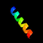
| 1 |
|
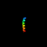
PDB 3viq chain C
Region: 44 - 66
Aligned: 23
Modelled: 23
Confidence: 22.1%
Identity: 13%
PDB header:recombination activator
Chain: C: PDB Molecule:swi5-dependent recombination dna repair protein 1;
PDBTitle: crystal structure of swi5-sfr1 complex from fission yeast
Phyre2
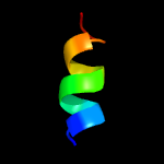
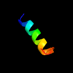
| 2 |
|
PDB 4etr chain A
Region: 59 - 104
Aligned: 46
Modelled: 46
Confidence: 21.6%
Identity: 22%
PDB header:unknown function
Chain: A: PDB Molecule:putative uncharacterized protein;
PDBTitle: x-ray structure of pa2169 from pseudomonas aeruginosa
Phyre2
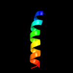
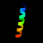
| 3 |
|
PDB 6fo2 chain R
Region: 14 - 36
Aligned: 23
Modelled: 23
Confidence: 10.7%
Identity: 26%
PDB header:membrane protein
Chain: R: PDB Molecule:cytochrome b-c1 complex subunit rieske, mitochondrial;
PDBTitle: cryoem structure of bovine cytochrome bc1 with no ligand bound
Phyre2
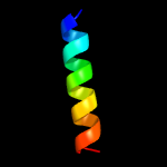
| 4 |
|
PDB 1xnl chain A
Region: 90 - 103
Aligned: 13
Modelled: 14
Confidence: 10.7%
Identity: 54%
PDB header:viral protein
Chain: A: PDB Molecule:membrane protein gp37;
PDBTitle: aslv fusion peptide
Phyre2
| 5 |
|
PDB 2fyu chain E
Region: 16 - 36
Aligned: 21
Modelled: 21
Confidence: 10.0%
Identity: 29%
PDB header:oxidoreductase
Chain: E: PDB Molecule:ubiquinol-cytochrome c reductase iron-sulfur subunit,
PDBTitle: crystal structure of bovine heart mitochondrial bc1 with jg1442 inhibitor
Phyre2
| 6 |
|
PDB 3vep chain X
Region: 44 - 62
Aligned: 19
Modelled: 19
Confidence: 10.0%
Identity: 32%
PDB header:membrane protein/transcription
Chain: X: PDB Molecule:uncharacterized protein rv3413c/mt3522;
PDBTitle: crystal structure of sigd4 in complex with its negative regulator rsda
Phyre2
| 7 |
|
PDB 3vep chain C
Region: 44 - 62
Aligned: 19
Modelled: 19
Confidence: 9.8%
Identity: 32%
PDB header:membrane protein/transcription
Chain: C: PDB Molecule:uncharacterized protein rv3413c/mt3522;
PDBTitle: crystal structure of sigd4 in complex with its negative regulator rsda
Phyre2
| 8 |
|
PDB 3no7 chain A
Region: 92 - 109
Aligned: 18
Modelled: 18
Confidence: 7.8%
Identity: 28%
PDB header:dna binding protein
Chain: A: PDB Molecule:putative plasmid related protein;
PDBTitle: crystal structure of the centromere-binding protein parb from plasmid2 pcxc100
Phyre2
| 9 |
|
PDB 2fyn chain O
Region: 17 - 36
Aligned: 20
Modelled: 20
Confidence: 6.7%
Identity: 30%
PDB header:oxidoreductase
Chain: O: PDB Molecule:ubiquinol-cytochrome c reductase iron-sulfur
PDBTitle: crystal structure analysis of the double mutant rhodobacter2 sphaeroides bc1 complex
Phyre2
| 10 |
|
PDB 3he5 chain D
Region: 95 - 102
Aligned: 8
Modelled: 8
Confidence: 5.1%
Identity: 63%
PDB header:de novo protein
Chain: D: PDB Molecule:synzip2;
PDBTitle: heterospecific coiled-coil pair synzip2:synzip1
Phyre2